WB
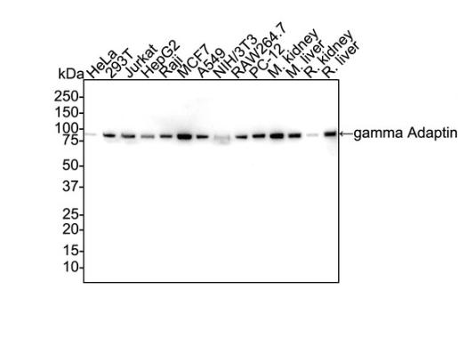
Western blot analysis of gamma Adaptin on different lysates with Rabbit anti-gamma Adaptin antibody at 1/1,000 dilution. Lane 1: HeLa cell lysate (20 µg/Lane) Lane 2: 293T cell lysate (20 µg/Lane) Lane 3: Jurkat cell lysate (20 µg/Lane) Lane 4: HepG2 cell lysate (20 µg/Lane) Lane 5: Raji cell lysate (20 µg/Lane) Lane 6: MCF7 cell lysate (20 µg/Lane) Lane 7: A549 cell lysate (20 µg/Lane) Lane 8: NIH/3T3 cell lysate (20 µg/Lane) Lane 9: RAW264.7 cell lysate (20 µg/Lane) Lane 10: PC-12 cell lysate (20 µg/Lane) Lane 11: Mouse kidney tissue lysate (40 µg/Lane) Lane 12: Mouse liver tissue lysate (40 µg/Lane) Lane 13: Rat kidney tissue lysate (40 µg/Lane) Lane 14: Rat liver tissue lysate (40 µg/Lane) Exposure time: 1 minute 46 seconds; 4-20% SDS-PAGE gel. Proteins were transferred to a PVDF membrane and blocked with 5% NFDM/TBST for 1 hour at room temperature. The primary antibody at 1/1,000 dilution was used in 5% NFDM/TBST at 4℃ overnight. Goat Anti-Rabbit IgG - HRP Secondary Antibody at 1/50,000 dilution was used for 1 hour at room temperature.WB
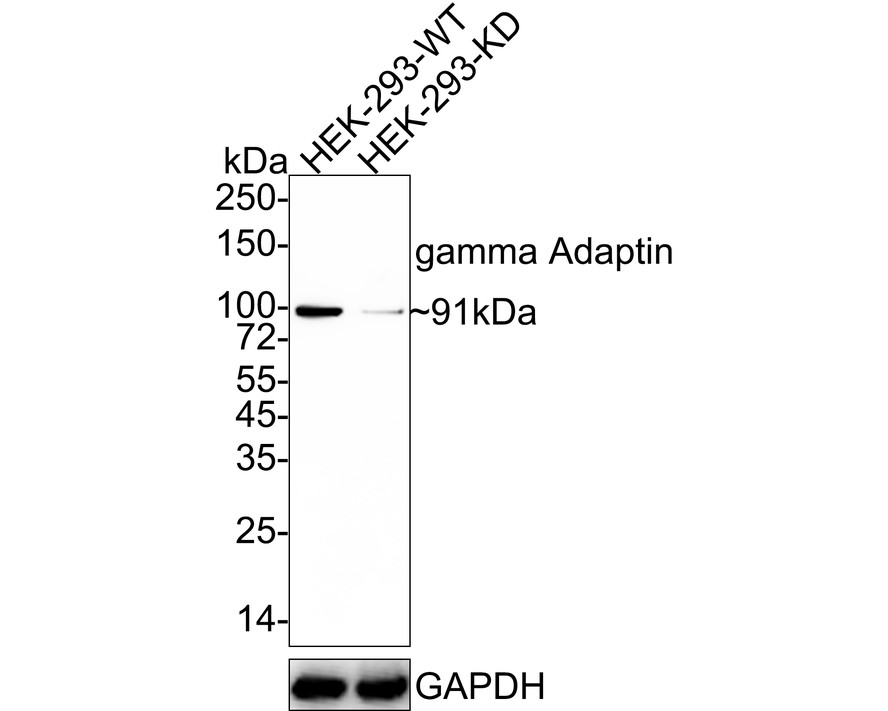
Western blot analysis of gamma Adaptin on different lysates with Rabbit anti-gamma Adaptin antibody at 1/1,000 dilution. Lane 1: HEK-293-si NT cell lysate, Lane 2: HEK-293-si gamma Adaptin cell lysate, Lysates/proteins at 10 µg/Lane. Exposure time: 2 minutes 18 seconds; 4-20% SDS-PAGE gel. Proteins were transferred to a PVDF membrane and blocked with 5% NFDM/TBST for 1 hour at room temperature. The primary antibody at 1/1,000 dilution was used in 5% NFDM/TBST at 4℃ overnight. Goat Anti-Rabbit IgG - HRP Secondary Antibody at 1/50,000 dilution was used for 1 hour at room temperature.IHC
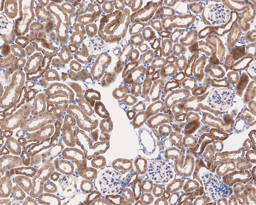
Immunohistochemical analysis of paraffin-embedded mouse kidney tissue with Rabbit anti-gamma Adaptin antibody at 1/200 dilution. The section was pre-treated using heat mediated antigen retrieval with Tris-EDTA buffer (pH 9.0) for 20 minutes. The tissues were blocked in 1% BSA for 20 minutes at room temperature, washed with ddH2O and PBS, and then probed with the primary antibody at 1/200 dilution for 1 hour at room temperature. The detection was performed using an HRP conjugated compact polymer system. DAB was used as the chromogen. Tissues were counterstained with hematoxylin and mounted with DPX.IHC
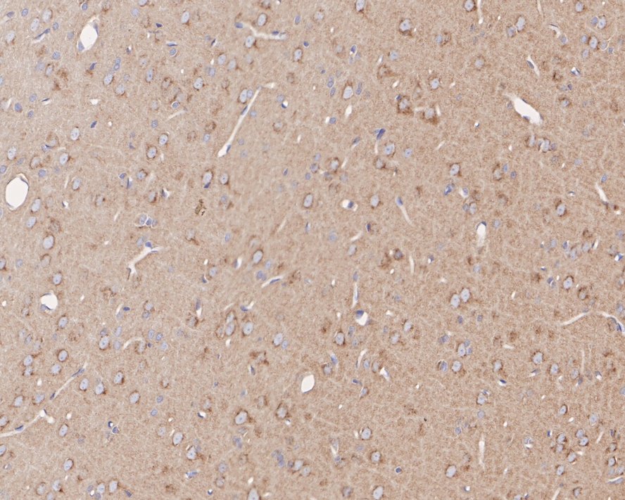
Immunohistochemical analysis of paraffin-embedded rat brain tissue with Rabbit anti-gamma Adaptin antibody at 1/200 dilution. The section was pre-treated using heat mediated antigen retrieval with Tris-EDTA buffer (pH 9.0) for 20 minutes. The tissues were blocked in 1% BSA for 20 minutes at room temperature, washed with ddH2O and PBS, and then probed with the primary antibody at 1/200 dilution for 1 hour at room temperature. The detection was performed using an HRP conjugated compact polymer system. DAB was used as the chromogen. Tissues were counterstained with hematoxylin and mounted with DPX.| Product Name | gamma Adaptin Recombinant Rabbit Monoclonal Antibody |
|---|---|
| Antibody Type | Primary Antibodies |
| Immunogen | Recombinant protein within human gamma Adaptin aa 623-822 / 822. |
| Clonality | Monoclonal |
|---|---|
| Isotype | IgG |
| Host Species | Rabbit |
| Tested Applications | IHCWB |
| WB:1:1000 IHC:1:200 |
|
| Species Reactivity | HumanMouseRat |
| Concentration | 1mg/ml |
| Purification | Protein A |
| Gene Symbol | AP1G1 |
|---|---|
| Gene Synonyms | ADTG CLAPG1 USRISD |
| Gene Full Name | adaptor related protein complex 1 subunit gamma 1 |
| Gene Summary | Adaptins are important components of clathrin-coated vesicles transporting ligand-receptor complexes from the plasma membrane or from the trans-Golgi network to lysosomes. The adaptin family of proteins is composed of four classes of molecules named alpha, beta-, beta prime- and gamma- adaptins. Adaptins, together with medium and small subunits, form a heterotetrameric complex called an adaptor, whose role is to promote the formation of clathrin-coated pits and vesicles. The protein encoded by this gene is a gamma-adaptin protein and it belongs to the adaptor complexes large subunits family. Two transcript variants encoding different isoforms have been found for this gene. [provided by RefSeq, Jul 2008] |
| Alternative Names | Adapter-related protein complex 1 subunit gamma-1 antibody Adaptor protein complex AP 1 gamma 1 subunit antibody Adaptor protein complex AP 1 subunit gamma 1 antibody Adaptor protein complex AP-1 subunit gamma-1 antibody Adaptor related protein complex 1 gamma 1 subunit antibody ADTG antibody AP 1 complex subunit gamma 1 antibody AP-1 complex subunit gamma-1 antibody AP1G1 antibody AP1G1_HUMAN antibody Adapter-related protein complex 1 subunit gamma-1 antibody Adaptor protein complex AP 1 gamma 1 subunit antibody Adaptor protein complex AP 1 subunit gamma 1 antibody Adaptor protein complex AP-1 subunit gamma-1 antibody Adaptor related protein complex 1 gamma 1 subunit antibody ADTG antibody AP 1 complex subunit gamma 1 antibody AP-1 complex subunit gamma-1 antibody AP1G1 antibody AP1G1_HUMAN antibody CLAPG1 antibody Clathrin assembly protein complex 1 gamma 1 large chain antibody Clathrin assembly protein complex 1 gamma large chain antibody Clathrin assembly protein complex 1 gamma-1 large chain antibody Clathrin associated/assembly/adaptor protein large gamma 1 antibody Gamma1 adaptin antibody Gamma1-adaptin antibody Golgi adaptor HA1/AP1 adaptin gamma subunit antibody Golgi adaptor HA1/AP1 adaptin subunit gamma 1 antibody Golgi adaptor HA1/AP1 adaptin subunit gamma-1 antibody MGC18255 antibody |
| Molecular Weight(MW) | 91 kDa |
| Cellular Localization | Golgi apparatus, Cytoplasmic vesicle, clathrin-coated vesicle membrane, Cytoplasm, perinuclear region, |
WB
Western blot analysis of gamma Adaptin on different lysates with Rabbit anti-gamma Adaptin antibody at 1/1,000 dilution. Lane 1: HeLa cell lysate (20 µg/Lane) Lane 2: 293T cell lysate (20 µg/Lane) Lane 3: Jurkat cell lysate (20 µg/Lane) Lane 4: HepG2 cell lysate (20 µg/Lane) Lane 5: Raji cell lysate (20 µg/Lane) Lane 6: MCF7 cell lysate (20 µg/Lane) Lane 7: A549 cell lysate (20 µg/Lane) Lane 8: NIH/3T3 cell lysate (20 µg/Lane) Lane 9: RAW264.7 cell lysate (20 µg/Lane) Lane 10: PC-12 cell lysate (20 µg/Lane) Lane 11: Mouse kidney tissue lysate (40 µg/Lane) Lane 12: Mouse liver tissue lysate (40 µg/Lane) Lane 13: Rat kidney tissue lysate (40 µg/Lane) Lane 14: Rat liver tissue lysate (40 µg/Lane) Exposure time: 1 minute 46 seconds; 4-20% SDS-PAGE gel. Proteins were transferred to a PVDF membrane and blocked with 5% NFDM/TBST for 1 hour at room temperature. The primary antibody at 1/1,000 dilution was used in 5% NFDM/TBST at 4℃ overnight. Goat Anti-Rabbit IgG - HRP Secondary Antibody at 1/50,000 dilution was used for 1 hour at room temperature.
WB
Western blot analysis of gamma Adaptin on different lysates with Rabbit anti-gamma Adaptin antibody at 1/1,000 dilution. Lane 1: HEK-293-si NT cell lysate, Lane 2: HEK-293-si gamma Adaptin cell lysate, Lysates/proteins at 10 µg/Lane. Exposure time: 2 minutes 18 seconds; 4-20% SDS-PAGE gel. Proteins were transferred to a PVDF membrane and blocked with 5% NFDM/TBST for 1 hour at room temperature. The primary antibody at 1/1,000 dilution was used in 5% NFDM/TBST at 4℃ overnight. Goat Anti-Rabbit IgG - HRP Secondary Antibody at 1/50,000 dilution was used for 1 hour at room temperature.
IHC
Immunohistochemical analysis of paraffin-embedded mouse kidney tissue with Rabbit anti-gamma Adaptin antibody at 1/200 dilution. The section was pre-treated using heat mediated antigen retrieval with Tris-EDTA buffer (pH 9.0) for 20 minutes. The tissues were blocked in 1% BSA for 20 minutes at room temperature, washed with ddH2O and PBS, and then probed with the primary antibody at 1/200 dilution for 1 hour at room temperature. The detection was performed using an HRP conjugated compact polymer system. DAB was used as the chromogen. Tissues were counterstained with hematoxylin and mounted with DPX.
IHC
Immunohistochemical analysis of paraffin-embedded rat brain tissue with Rabbit anti-gamma Adaptin antibody at 1/200 dilution. The section was pre-treated using heat mediated antigen retrieval with Tris-EDTA buffer (pH 9.0) for 20 minutes. The tissues were blocked in 1% BSA for 20 minutes at room temperature, washed with ddH2O and PBS, and then probed with the primary antibody at 1/200 dilution for 1 hour at room temperature. The detection was performed using an HRP conjugated compact polymer system. DAB was used as the chromogen. Tissues were counterstained with hematoxylin and mounted with DPX.| Application Notes | WB:1:1000 IHC:1:200 |
|---|
| Form | Liquid |
|---|---|
| Storage Instructions | Store at +4℃ after thawing. Aliquot store at -20℃. Avoid repeated freeze / thaw cycles. |
| Storage Buffer | PBS (pH7.4), 0.1% BSA, 40% Glycerol. Preservative: 0.05% Sodium Azide. |


