
WB
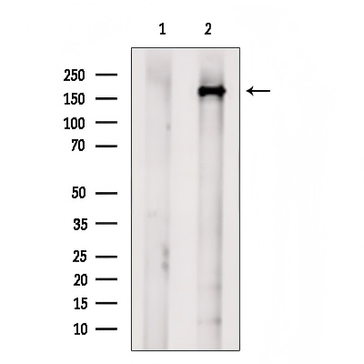
Western blot analysis of extracts from RAW264.7 cells, using USP54 Antibody. The lane on the left was treated with blocking peptide.IHC
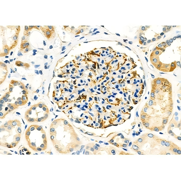
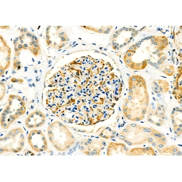
USP54 Antibody at 1/100 staining Human kidney cancer and adjacent normal tissues by IHC-P. The sample was formaldehyde fixed and a heat mediated antigen retrieval step in citrate buffer was performed. The sample was then blocked and incubated with the primary antibody at 4°C overnight. An HRP conjugated anti-Rabbit antibody was used as the secondary antibody.IHC
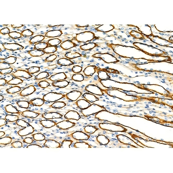
USP54 Antibody at 1/100 staining Mouse kidney tissue by IHC-P. The sample was formaldehyde fixed and a heat mediated antigen retrieval step in citrate buffer was performed. The sample was then blocked and incubated with the primary antibody at 4°C overnight. An HRP conjugated anti-Rabbit antibody was used as the secondary antibody.IHC
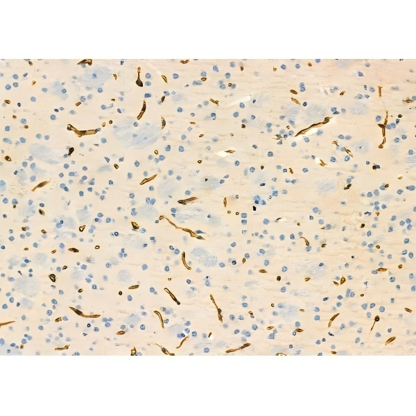
USP54 Antibody at 1/100 staining Mouse brain tissue by IHC-P. The sample was formaldehyde fixed and a heat mediated antigen retrieval step in citrate buffer was performed. The sample was then blocked and incubated with the primary antibody at 4°C overnight. An HRP conjugated anti-Rabbit antibody was used as the secondary antibody.IHC
USP54 Antibody at 1/100 staining Human pancreatic cancer and adjacent nomal tissues by IHC-P. The sample was formaldehyde fixed and a heat mediated antigen retrieval step in citrate buffer was performed. The sample was then blocked and incubated with the primary antibody at 4°C overnight. An HRP conjugated anti-Rabbit antibody was used as the secondary antibody.IHC
USP54 Antibody at 1/100 staining Human kidney cancer and adjacent normal tissues by IHC-P. The sample was formaldehyde fixed and a heat mediated antigen retrieval step in citrate buffer was performed. The sample was then blocked and incubated with the primary antibody at 4°C overnight. An HRP conjugated anti-Rabbit antibody was used as the secondary antibody.WB
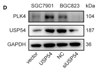
5 USP54 combines with PLK4 and mediates its deubiquitination (A) Microarray analysis of mRNAs extracted from SGC7901-vector and SGC7901-CEP120 cells. (B) Results of the ubiquitination level of PLK4 in HEK293T cells, which were co-transfected with Flag-PLK4, HA-Ub, and one of the three DUBs. (C) WB results of USP54 and PLK4 expression in GC tissues. p value derived from Pearson correlation analysis. (D) WB results of PLK4 and USP54 after USP54 overexpression or knockdown. (E) CoIP analysis of the interaction between PLK4 and USP54 in SGC7901 and BGC823 cells. (F) HEK 293T cells were co-transfected with His-USP54 and Flag-PLK4 full length or Flag-PLK4 (1-740), Flag-PLK4 (1-270), and Flag-PLK4 (270-890) constructs and harvested for WB and IP analysis. (G) HEK 293T cells were co-transfected with His-USP54 and Flag-PLK4 wildtype or Flag-PLK4 lysine residue mutants and harvested for IP analysis. (H) Ubiquitination level of PLK4 after USP54 overexpression or knockdown examined by ubiquitination assay. (I) WB results of PLK4 in SGC7901 cells transfected with Lenti-CEP120 and siUSP54 and BGC823 cells transfected with Lenti-shCEP120 and USP54 plasmid.| Product Name | USP54 Antibody |
|---|---|
| Antibody Type | Primary Antibodies |
| Immunogen | A synthesized peptide derived from human USP54, corresponding to a region within the internal amino acids. |
| Clonality | Polyclonal |
|---|---|
| Isotype | IgG |
| Host Species | Rabbit |
| Tested Applications | IHCWB |
| WB:1:1000-3000 IHC:1:50-1:200 |
|
| Species Reactivity | HumanMouseRat |
| Concentration | 1mg/ml |
| Purification | Affinity purified |
| Gene Symbol | USP54 |
|---|---|
| Gene Synonyms | C10orf29 bA137L10.3 bA137L10.4 |
| Gene Full Name | ubiquitin specific peptidase 54 |
| Gene Summary | Enables K63-linked deubiquitinase activity and cysteine-type deubiquitinase activity. Predicted to be involved in protein deubiquitination and proteolysis. [provided by Alliance of Genome Resources, Jul 2025] |
| Alternative Names | USP54 bA137L10.3 bA137L10.4 C10orf29 FLJ37318 Inactive ubiquitin carboxyl-terminal hydrolase 54 nactive ubiquitin-specific peptidase 54 |
| Molecular Weight(MW) | 187kDa; 187kD(Calculated). |
| Entrez Gene | 159195 |
|---|
WB
Western blot analysis of extracts from RAW264.7 cells, using USP54 Antibody. The lane on the left was treated with blocking peptide.
IHC
USP54 Antibody at 1/100 staining Human kidney cancer and adjacent normal tissues by IHC-P. The sample was formaldehyde fixed and a heat mediated antigen retrieval step in citrate buffer was performed. The sample was then blocked and incubated with the primary antibody at 4°C overnight. An HRP conjugated anti-Rabbit antibody was used as the secondary antibody.
IHC
USP54 Antibody at 1/100 staining Mouse kidney tissue by IHC-P. The sample was formaldehyde fixed and a heat mediated antigen retrieval step in citrate buffer was performed. The sample was then blocked and incubated with the primary antibody at 4°C overnight. An HRP conjugated anti-Rabbit antibody was used as the secondary antibody.
IHC
USP54 Antibody at 1/100 staining Mouse brain tissue by IHC-P. The sample was formaldehyde fixed and a heat mediated antigen retrieval step in citrate buffer was performed. The sample was then blocked and incubated with the primary antibody at 4°C overnight. An HRP conjugated anti-Rabbit antibody was used as the secondary antibody.
IHC
USP54 Antibody at 1/100 staining Human pancreatic cancer and adjacent nomal tissues by IHC-P. The sample was formaldehyde fixed and a heat mediated antigen retrieval step in citrate buffer was performed. The sample was then blocked and incubated with the primary antibody at 4°C overnight. An HRP conjugated anti-Rabbit antibody was used as the secondary antibody.
IHC
USP54 Antibody at 1/100 staining Human kidney cancer and adjacent normal tissues by IHC-P. The sample was formaldehyde fixed and a heat mediated antigen retrieval step in citrate buffer was performed. The sample was then blocked and incubated with the primary antibody at 4°C overnight. An HRP conjugated anti-Rabbit antibody was used as the secondary antibody.
WB
5 USP54 combines with PLK4 and mediates its deubiquitination (A) Microarray analysis of mRNAs extracted from SGC7901-vector and SGC7901-CEP120 cells. (B) Results of the ubiquitination level of PLK4 in HEK293T cells, which were co-transfected with Flag-PLK4, HA-Ub, and one of the three DUBs. (C) WB results of USP54 and PLK4 expression in GC tissues. p value derived from Pearson correlation analysis. (D) WB results of PLK4 and USP54 after USP54 overexpression or knockdown. (E) CoIP analysis of the interaction between PLK4 and USP54 in SGC7901 and BGC823 cells. (F) HEK 293T cells were co-transfected with His-USP54 and Flag-PLK4 full length or Flag-PLK4 (1-740), Flag-PLK4 (1-270), and Flag-PLK4 (270-890) constructs and harvested for WB and IP analysis. (G) HEK 293T cells were co-transfected with His-USP54 and Flag-PLK4 wildtype or Flag-PLK4 lysine residue mutants and harvested for IP analysis. (H) Ubiquitination level of PLK4 after USP54 overexpression or knockdown examined by ubiquitination assay. (I) WB results of PLK4 in SGC7901 cells transfected with Lenti-CEP120 and siUSP54 and BGC823 cells transfected with Lenti-shCEP120 and USP54 plasmid.| Application Notes | WB:1:1000-3000 IHC:1:50-1:200 |
|---|
| Form | Liquid |
|---|---|
| Storage Instructions | Store at -20 °C. Stable for 12 months from date of receipt. |
| Storage Buffer | Rabbit IgG in phosphate buffered saline , pH 7.4, 150mM NaCl, 0.02% sodium azide and 50% glycerol. |


