WB
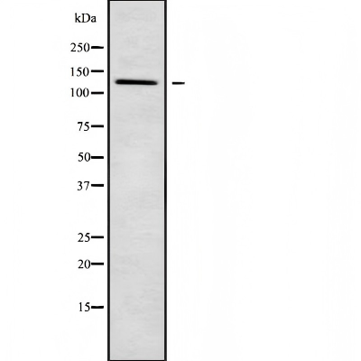
Western blot analysis of PDE3B using HuvEc whole cell lysatesIHC
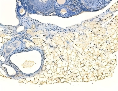
PDE3B Antibody at 1/100 staining rat muscle tissue by IHC-P. The sample was formaldehyde fixed and a heat mediated antigen retrieval step in citrate buffer was performed. The sample was then blocked and incubated with the primary antibody at 4°C overnight. An HRP conjugated anti-rabbit antibody was used as the secondary antibody.ICC/IF
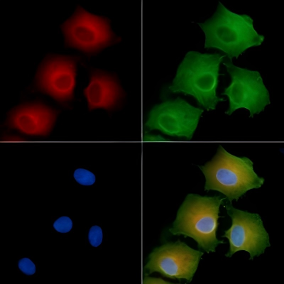
PDE3B Antibody staining A549 cells by IF/ICC. The samples were fixed with PFA and permeabilized in 0.1% Triton X-100, then blocked in 10% serum for 45 minutes at 25°C. Samples were then incubated with primary Ab(#DF9372) and mouse anti-beta tubulin Ab(#T0023) for 1 hour at 37°C. An AlexaFluor594 conjugated goat anti-rabbit IgG Ab(Red) and an AlexaFluor488 conjugated goat anti-mouse IgG Ab(Green) were used as the secondary antibody. The nuclear counter stain is DAPI (blue).| Product Name | PDE3B Antibody |
|---|---|
| Antibody Type | Primary Antibodies |
| Immunogen | A synthesized peptide derived from human PDE3B, corresponding to a region within the internal amino acids. |
| Clonality | Polyclonal |
|---|---|
| Isotype | IgG |
| Host Species | Rabbit |
| Tested Applications | ICC/IFIHCWB |
| WB:1:1000-1:3000 IHC:1:50-1:200 ICC/IF:1:100-1:500 |
|
| Species Reactivity | Human |
| Concentration | 1mg/ml |
| Purification | Affinity purified |
| Gene Symbol | PDE3B |
|---|---|
| Gene Synonyms | HcGIP1 cGIPDE1 |
| Gene Full Name | phosphodiesterase 3B |
| Gene Summary | Enables 3',5'-cyclic-AMP phosphodiesterase activity. Involved in several processes, including negative regulation of angiogenesis; negative regulation of lipid catabolic process; and regulation of phosphatidylinositol 3-kinase/protein kinase B signal transduction. Located in membrane. Part of guanyl-nucleotide exchange factor complex. [provided by Alliance of Genome Resources, Jul 2025] |
| Alternative Names | 5''-cyclic phosphodiesterase B Adipocyte cAMP phosphodiesterase cGMP inhibited AI847709 cAMP Phosphodiesterase adipocyte cGMP-inhibited CGI-PDE B CGIP1 CGIPDE B CGIPDE1 cGMP inhibited 3 5 cyclic phosphodiesterase B CGMP inhibited cAMP phophodiesterase cGMP-inhibited 3'' Cyclic GMP inhibited phosphodiesterase B Cyclic GMP-inhibited phosphodiesterase B Cyclic nucleotide phosphodiesterase HcGIP1 HcGIP1 rat homolog of Pde3b PDE3B_HUMAN Phosphodiesterase 3B Phosphodiesterase 3B cGMP inhibited |
| Molecular Weight(MW) | 124kDa; 124kD(Calculated). |
| Cellular Localization | Membrane>Multi-pass membrane protein. |
WB
Western blot analysis of PDE3B using HuvEc whole cell lysates
IHC
PDE3B Antibody at 1/100 staining rat muscle tissue by IHC-P. The sample was formaldehyde fixed and a heat mediated antigen retrieval step in citrate buffer was performed. The sample was then blocked and incubated with the primary antibody at 4°C overnight. An HRP conjugated anti-rabbit antibody was used as the secondary antibody.
ICC/IF
PDE3B Antibody staining A549 cells by IF/ICC. The samples were fixed with PFA and permeabilized in 0.1% Triton X-100, then blocked in 10% serum for 45 minutes at 25°C. Samples were then incubated with primary Ab(#DF9372) and mouse anti-beta tubulin Ab(#T0023) for 1 hour at 37°C. An AlexaFluor594 conjugated goat anti-rabbit IgG Ab(Red) and an AlexaFluor488 conjugated goat anti-mouse IgG Ab(Green) were used as the secondary antibody. The nuclear counter stain is DAPI (blue).| Application Notes | WB:1:1000-1:3000 IHC:1:50-1:200 ICC/IF:1:100-1:500 |
|---|
| Form | Liquid |
|---|---|
| Storage Instructions | Store at -20 °C. Stable for 12 months from date of receipt. |
| Storage Buffer | Rabbit IgG in phosphate buffered saline , pH 7.4, 150mM NaCl, 0.02% sodium azide and 50% glycerol. |


