WB
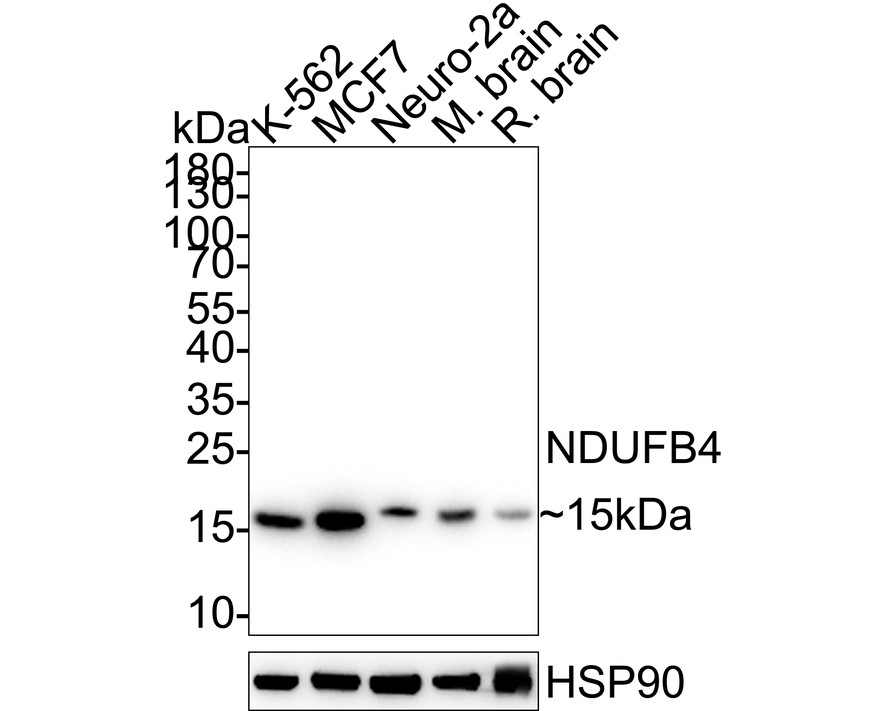
Western blot analysis of NDUFB4 on different lysates with Rabbit anti-NDUFB4 antibody at 1/2,000 dilution. Lane 1: K-562 cell lysate, Lane 2: MCF7 cell lysate, Lane 3: Neuro-2a cell lysate, Lane 4: Mouse brain tissue lysate, Lane 5: Rat brain tissue lysate, Lysates/proteins at 20 µg/Lane. Exposure time: 30 seconds; ECL: K1801; 4-20% SDS-PAGE gel. Proteins were transferred to a PVDF membrane and blocked with 5% NFDM/TBST for 1 hour at room temperature. The primary antibody at 1/2,000 dilution was used in 5% NFDM/TBST at 4℃ overnight. Goat Anti-Rabbit IgG - HRP Secondary Antibody at 1/50,000 dilution was used for 1 hour at room temperature.WB
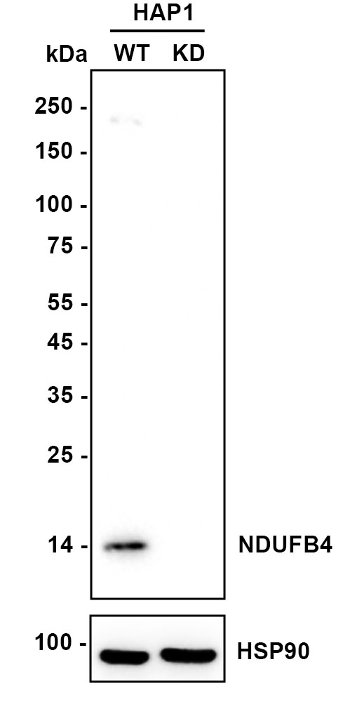
Western blot analysis of NDUFB4 on different lysates with Rabbit anti-NDUFB4 antibody at 1/1,000 dilution. Lane 1: HAP1-parental cell lysate, Lane 2: HAP1-NDUFB4 KD cell lysate, Lysates/proteins at 10 µg/Lane. Exposure time: 20 seconds; ECL: K1801; 4-20% SDS-PAGE gel. Proteins were transferred to a PVDF membrane and blocked with 5% NFDM/TBST for 1 hour at room temperature. The primary antibody at 1/1,000 dilution was used in K1803 at 4℃ overnight. Goat Anti-Rabbit IgG - HRP Secondary Antibody at 1/50,000 dilution was used for 1 hour at room temperature.IHC
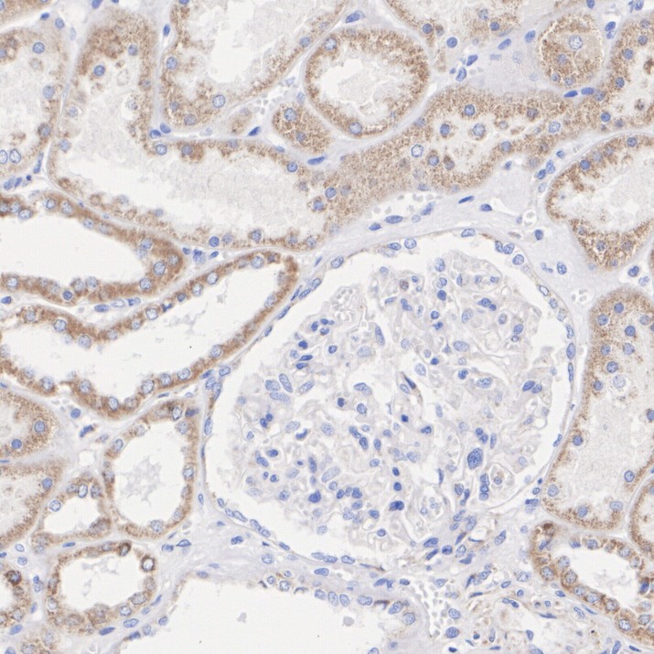
Immunohistochemical analysis of paraffin-embedded human kidney tissue with Rabbit anti-NDUFB4 antibody at 1/1,000 dilution. The section was pre-treated using heat mediated antigen retrieval with Tris-EDTA buffer (pH 9.0) for 20 minutes. The tissues were blocked in 1% BSA for 20 minutes at room temperature, washed with ddH2O and PBS, and then probed with the primary antibody at 1/1,000 dilution for 1 hour at room temperature. The detection was performed using an HRP conjugated compact polymer system. DAB was used as the chromogen. Tissues were counterstained with hematoxylin and mounted with DPX.| Product Name | NDUFB4 Recombinant Rabbit Monoclonal Antibody |
|---|---|
| Antibody Type | Primary Antibodies |
| Immunogen | Recombinant protein within human NDUFB4 aa 1-129/129. |
| Clonality | Monoclonal |
|---|---|
| Isotype | IgG |
| Host Species | Rabbit |
| Tested Applications | IHCWB |
| WB:1:1,000-1:2,000 IHC:1:1,000 |
|
| Species Reactivity | HumanMouseRat |
| Concentration | 1mg/ml |
| Purification | Protein A |
| Gene Symbol | MYL12B |
|---|---|
| Gene Synonyms | B15 antibody,CI-B15 antibody,Complex I B15 subunit antibody,Complex I-B15 antibody,NADH dehydrogenase 1 beta subcomplex 4 15kDa antibody,NADH dehydrogenase 1 beta subcomplex subunit 4 antibody,NADH ubiquinone oxidoreductase B15 subunit antibody,NADH-ubiquinone oxidoreductase B15 subunit antibody,NDUB4_HUMAN antibody,NDUFB4 antibody |
| Gene Full Name | NADH:ubiquinone oxidoreductase subunit B4 |
| Gene Summary | The activity of nonmuscle myosin II (see MYH9; MIM 160775) is regulated by phosphorylation of a regulatory light chain, such as MRLC2. This phosphorylation results in higher MgATPase activity and the assembly of myosin II filaments (Iwasaki et al., 2001 [PubMed 11942626]).[supplied by OMIM, Mar 2008] |
| Alternative Names | B15 antibody CI-B15 antibody Complex I B15 subunit antibody Complex I-B15 antibody NADH dehydrogenase (ubiquinone) 1 beta subcomplex 4 15kDa antibody NADH dehydrogenase [ubiquinone] 1 beta subcomplex subunit 4 antibody NADH ubiquinone oxidoreductase B15 subunit antibody NADH-ubiquinone oxidoreductase B15 subunit antibody NDUB4_HUMAN antibody NDUFB4 antibody |
| Molecular Weight(MW) | 15 kDa |
| Cellular Localization | Mitochondrion inner membrane. |
WB
Western blot analysis of NDUFB4 on different lysates with Rabbit anti-NDUFB4 antibody at 1/2,000 dilution. Lane 1: K-562 cell lysate, Lane 2: MCF7 cell lysate, Lane 3: Neuro-2a cell lysate, Lane 4: Mouse brain tissue lysate, Lane 5: Rat brain tissue lysate, Lysates/proteins at 20 µg/Lane. Exposure time: 30 seconds; ECL: K1801; 4-20% SDS-PAGE gel. Proteins were transferred to a PVDF membrane and blocked with 5% NFDM/TBST for 1 hour at room temperature. The primary antibody at 1/2,000 dilution was used in 5% NFDM/TBST at 4℃ overnight. Goat Anti-Rabbit IgG - HRP Secondary Antibody at 1/50,000 dilution was used for 1 hour at room temperature.
WB
Western blot analysis of NDUFB4 on different lysates with Rabbit anti-NDUFB4 antibody at 1/1,000 dilution. Lane 1: HAP1-parental cell lysate, Lane 2: HAP1-NDUFB4 KD cell lysate, Lysates/proteins at 10 µg/Lane. Exposure time: 20 seconds; ECL: K1801; 4-20% SDS-PAGE gel. Proteins were transferred to a PVDF membrane and blocked with 5% NFDM/TBST for 1 hour at room temperature. The primary antibody at 1/1,000 dilution was used in K1803 at 4℃ overnight. Goat Anti-Rabbit IgG - HRP Secondary Antibody at 1/50,000 dilution was used for 1 hour at room temperature.
IHC
Immunohistochemical analysis of paraffin-embedded human kidney tissue with Rabbit anti-NDUFB4 antibody at 1/1,000 dilution. The section was pre-treated using heat mediated antigen retrieval with Tris-EDTA buffer (pH 9.0) for 20 minutes. The tissues were blocked in 1% BSA for 20 minutes at room temperature, washed with ddH2O and PBS, and then probed with the primary antibody at 1/1,000 dilution for 1 hour at room temperature. The detection was performed using an HRP conjugated compact polymer system. DAB was used as the chromogen. Tissues were counterstained with hematoxylin and mounted with DPX.| Application Notes | WB:1:1,000-1:2,000 IHC:1:1,000 |
|---|
| Form | Liquid |
|---|---|
| Storage Instructions | Shipped at 4℃. Store at +4℃ short term (1-2 weeks). It is recommended to aliquot into single-use upon delivery. Store at -20℃ long term. |
| Storage Buffer | PBS (pH7.4), 0.1% BSA, 40% Glycerol. Preservative: 0.05% Sodium Azide. |


