WB
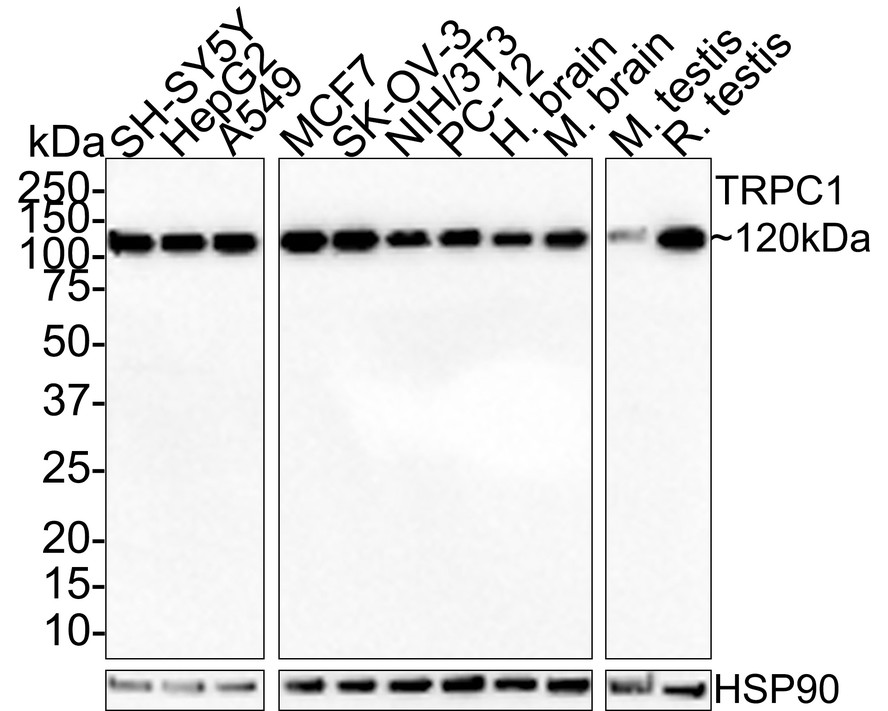
Western blot analysis of TRPC1 on different lysates with Rabbit anti-TRPC1 antibody at 1/1,000 dilution. Lane 1: SH-SY5Y cell lysate 15 µg/Lane), Lane 2: HepG2 cell lysate 15 µg/Lane), Lane 3: A549 cell lysate 15 µg/Lane), Lane 4: MCF7 cell lysate 15 µg/Lane), Lane 5: SK-OV-3 cell lysate 15 µg/Lane), Lane 6: NIH/3T3 cell lysate 15 µg/Lane), Lane 7: PC-12 cell lysate 15 µg/Lane), Lane 8: Human brain tissue lysate (30 µg/Lane), Lane 9: Mosue brain tissue lysate (30 µg/Lane), Lane 10: Mosue testis tissue lysate (30 µg/Lane), Lane 11: Rat testis tissue lysate (30 µg/Lane), Exposure time: 3 minutes; 4-20% SDS-PAGE gel. Proteins were transferred to a PVDF membrane and blocked with 5% NFDM/TBST for 1 hour at room temperature. The primary antibody at 1/1,000 dilution was used in 5% NFDM/TBST at room temperature for 2 hours. Goat Anti-Rabbit IgG - HRP Secondary Antibody at 1:100,000 dilution was used for 1 hour at room temperature.| Product Name | TRPC1 Recombinant Rabbit Monoclonal Antibody |
|---|---|
| Antibody Type | Primary Antibodies |
| Immunogen | Recombinant protein within Human TRPC1 aa 408-507 / 793. |
| Clonality | Monoclonal |
|---|---|
| Isotype | IgG |
| Host Species | Rabbit |
| Tested Applications | WB |
| WB:1:1000-1:2000 |
|
| Species Reactivity | HumanMouseRat |
| Concentration | 1mg/ml |
| Purification | Protein A |
| Gene Symbol | TRPC1 |
|---|---|
| Gene Synonyms | TRP1 HTRP-1 |
| Gene Full Name | transient receptor potential cation channel subfamily C member 1 |
| Gene Summary | The protein encoded by this gene is a membrane protein that can form a non-selective channel permeable to calcium and other cations. The encoded protein appears to be induced to form channels by a receptor tyrosine kinase-activated phosphatidylinositol second messenger system and also by depletion of intracellular calcium stores. Two transcript variants encoding different isoforms have been found for this gene. [provided by RefSeq, Oct 2011] |
| Alternative Names | HTRP 1 antibody HTRP1 antibody MGC133334 antibody MGC133335 antibody Mtrp1 antibody Short transient receptor potential channel 1 antibody Transient receptor potential canonical 1 antibody Transient receptor potential cation channel subfamily C member 1 antibody Transient receptor potential channel 1 antibody Transient receptor protein 1 antibody HTRP 1 antibody HTRP1 antibody MGC133334 antibody MGC133335 antibody Mtrp1 antibody Short transient receptor potential channel 1 antibody Transient receptor potential canonical 1 antibody Transient receptor potential cation channel subfamily C member 1 antibody Transient receptor potential channel 1 antibody Transient receptor protein 1 antibody TRP 1 antibody TRP 1 protein antibody Trp related protein 1 antibody TRP-1 antibody TRP1 antibody TRP1 protein antibody TrpC1 antibody Trpc1 transient receptor potential cation channel subfamily C member 1 antibody TRPC1_HUMAN antibody Trrp1 antibody |
| Molecular Weight(MW) | 91 kDa |
| Cellular Localization | Membrane. |
WB
Western blot analysis of TRPC1 on different lysates with Rabbit anti-TRPC1 antibody at 1/1,000 dilution. Lane 1: SH-SY5Y cell lysate 15 µg/Lane), Lane 2: HepG2 cell lysate 15 µg/Lane), Lane 3: A549 cell lysate 15 µg/Lane), Lane 4: MCF7 cell lysate 15 µg/Lane), Lane 5: SK-OV-3 cell lysate 15 µg/Lane), Lane 6: NIH/3T3 cell lysate 15 µg/Lane), Lane 7: PC-12 cell lysate 15 µg/Lane), Lane 8: Human brain tissue lysate (30 µg/Lane), Lane 9: Mosue brain tissue lysate (30 µg/Lane), Lane 10: Mosue testis tissue lysate (30 µg/Lane), Lane 11: Rat testis tissue lysate (30 µg/Lane), Exposure time: 3 minutes; 4-20% SDS-PAGE gel. Proteins were transferred to a PVDF membrane and blocked with 5% NFDM/TBST for 1 hour at room temperature. The primary antibody at 1/1,000 dilution was used in 5% NFDM/TBST at room temperature for 2 hours. Goat Anti-Rabbit IgG - HRP Secondary Antibody at 1:100,000 dilution was used for 1 hour at room temperature.| Application Notes | WB:1:1000-1:2000 |
|---|
| Form | Liquid |
|---|---|
| Storage Instructions | Shipped at 4℃. Store at +4℃ short term (1-2 weeks). It is recommended to aliquot into single-use upon delivery. Store at -20℃ long term. |
| Storage Buffer | PBS (pH7.4), 0.1% BSA, 40% Glycerol. Preservative: 0.05% Sodium Azide. |


