WB
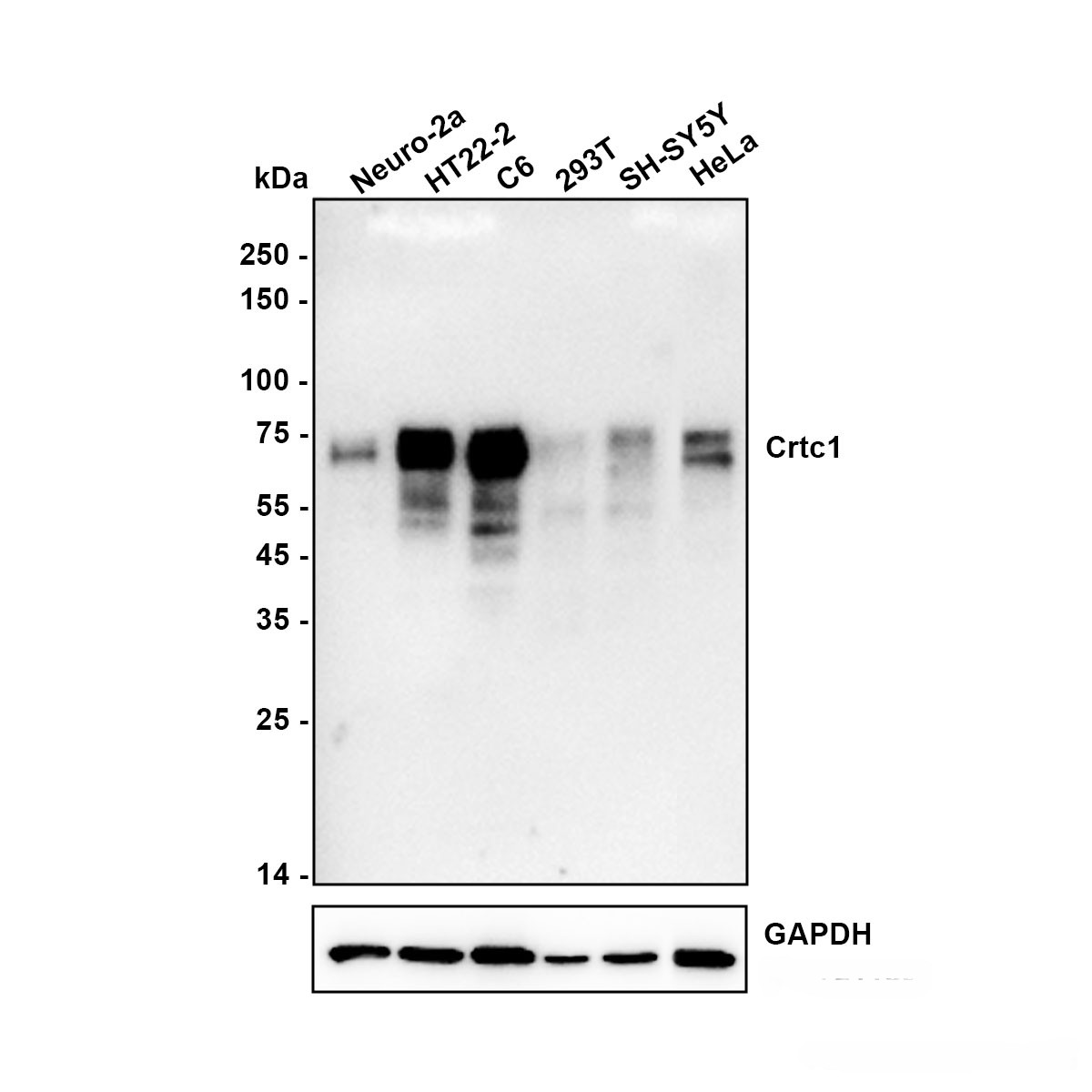
Western blot analysis of CRTC1 on different lysates with Rabbit anti-CRTC1 antibody at 1/5,000 dilution. Lane 1: Neuro-2a (Mouse brain neuroblastoma cell) cell lysate Lane 2: HT22 (Mouse hippocampal neuron cell) cell lysate Lane 3: C6 (Rat glioma cell) cell lysate Lane 4: 293T (Human embryonic kidney cell) cell lysate Lane 5: SH-SY5Y (Human neuroblastoma cell) cell lysate Lane 6: HeLa (Human cervical adenocarcinoma cell) cell lysate Lysates/proteins at 15 µg/Lane. Exposure time: 3 minutes; ECL: K1801WB
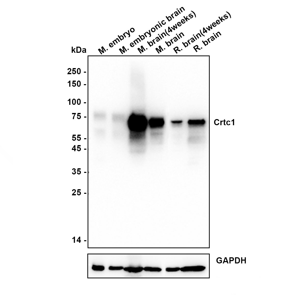
Western blot analysis of CRTC1 on different lysates with Rabbit anti-CRTC1 antibody at 1/5,000 dilution. Lane 1: Mouse embryo tissue lysate Lane 2: E14.0 mouse embryonic brain tissue lysate Lane 3: Mouse brain (4weeks) tissue lysate Lane 4: Mouse brain tissue lysate Lane 5: Rat brain (4weeks) tissue lysate Lane 6: Rat brain tissue lysate Lysates/proteins at 30 µg/Lane. Exposure time: 10 seconds; ECL: K1801FC
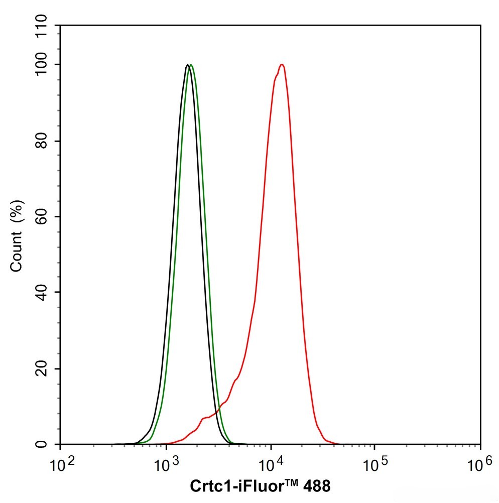
Application: Flow Cytometry (Intra) Species: Human Sample: 293T (Human embryonic kidney cells) Fixation: 4% Paraformaldehyde, 15 minutes at room temperature. Permeabilization: 0.1% Tween-20, 15 minutes at room temperature. Blocking: 1% BSA + 10% normal goat serum, 15 minutes at room temperature. Antibody dilution buffer: 1x PBS. Primary antibody:(1/1,000, Red) compared with Rabbit IgG Isotype Control (Green), 15 minutes at room temperature.FC
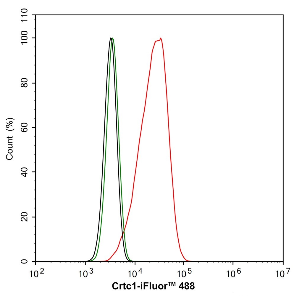
Application: Flow Cytometry (Intra) Species: Mouse Sample: Neuro-2a (Mouse brain neuroblastoma cells) Fixation: 4% Paraformaldehyde, 15 minutes at room temperature. Permeabilization: 0.1% Tween-20, 15 minutes at room temperature. Blocking: 1% BSA + 10% normal goat serum, 15 minutes at room temperature. Antibody dilution buffer: 1x PBS. Primary antibody:(1/1,000, Red) compared with Rabbit IgG Isotype Control (Green), 15 minutes at room temperature.IP
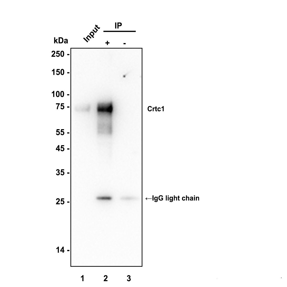
Immunoprecipitation (IP) CRTC1 was immunoprecipitated in 0.2 mg Neuro-2a (Mouse brain neuroblastoma cell) cell lysate with at 2 µg/10 µl beads. Western blot was performed from the immunoprecipitate using at 1/5,000 dilution. Anti-Rabbit IgG for IP Nano-secondary antibody (NBI01H) at 1/5,000 dilution was used for 1 hour at room temperature. Lane 1: Neuro-2A cell lysate (input) Lane 2: IP in Neuro-2A cell lysate Lane 3: Rabbit IgG instead of in Neuro-2A cell lysate Exposure time: 17 seconds Blocking: 5% NFDM/TBST, 1 hour at room temperature Predicted band size: 67 kDa Observed band size: 78 kDa| Product Name | CRTC1 Recombinant Rabbit Monoclonal Antibody |
|---|---|
| Antibody Type | Primary Antibodies |
| Immunogen | Recombinant protein within mouse CRTC1 aa 251-500. |
| Clonality | Monoclonal |
|---|---|
| Isotype | IgG |
| Host Species | Rabbit |
| Tested Applications | FCIPWB |
| WB:1:5000 FC1:1000IP1-2μg/sample |
|
| Species Reactivity | HumanMouseRat |
| Concentration | 1mg/ml |
| Purification | Protein A |
| Gene Symbol | CRTC1 |
|---|---|
| Gene Synonyms | MAML2 MECT1 Mam-2 TORC1 TORC-1 WAMTP1 |
| Gene Full Name | CREB regulated transcription coactivator 1 |
| Gene Summary | Enables cAMP response element binding protein binding activity. Involved in positive regulation of transcription by RNA polymerase II. Located in cytosol; nuclear body; and plasma membrane. [provided by Alliance of Genome Resources, Jul 2025] |
| Alternative Names | KIAA0616 antibody CREB regulated transcription coactivator 1 antibody CREB-regulated transcription coactivator 1 antibody CRTC1 antibody CRTC1_HUMAN antibody FLJ14027 antibody MECT 1 antibody Mucoepidermoid carcinoma translocated 1 antibody Mucoepidermoid carcinoma translocated protein 1 antibody TORC-1 antibody KIAA0616 antibody CREB regulated transcription coactivator 1 antibody CREB-regulated transcription coactivator 1 antibody CRTC1 antibody CRTC1_HUMAN antibody FLJ14027 antibody MECT 1 antibody Mucoepidermoid carcinoma translocated 1 antibody Mucoepidermoid carcinoma translocated protein 1 antibody TORC-1 antibody TORC1 antibody Transducer of CREB protein 1 antibody Transducer of regulated cAMP response element binding protein 1 antibody Transducer of regulated cAMP response element-binding protein (CREB) 1 antibody Transducer of regulated cAMP response element-binding protein 1 antibody WAMTP1 antibody |
| Molecular Weight(MW) | 67 kDa |
| Cellular Localization | Cytoplasm,Nucleus |
WB
Western blot analysis of CRTC1 on different lysates with Rabbit anti-CRTC1 antibody at 1/5,000 dilution. Lane 1: Neuro-2a (Mouse brain neuroblastoma cell) cell lysate Lane 2: HT22 (Mouse hippocampal neuron cell) cell lysate Lane 3: C6 (Rat glioma cell) cell lysate Lane 4: 293T (Human embryonic kidney cell) cell lysate Lane 5: SH-SY5Y (Human neuroblastoma cell) cell lysate Lane 6: HeLa (Human cervical adenocarcinoma cell) cell lysate Lysates/proteins at 15 µg/Lane. Exposure time: 3 minutes; ECL: K1801
WB
Western blot analysis of CRTC1 on different lysates with Rabbit anti-CRTC1 antibody at 1/5,000 dilution. Lane 1: Mouse embryo tissue lysate Lane 2: E14.0 mouse embryonic brain tissue lysate Lane 3: Mouse brain (4weeks) tissue lysate Lane 4: Mouse brain tissue lysate Lane 5: Rat brain (4weeks) tissue lysate Lane 6: Rat brain tissue lysate Lysates/proteins at 30 µg/Lane. Exposure time: 10 seconds; ECL: K1801
FC
Application: Flow Cytometry (Intra) Species: Human Sample: 293T (Human embryonic kidney cells) Fixation: 4% Paraformaldehyde, 15 minutes at room temperature. Permeabilization: 0.1% Tween-20, 15 minutes at room temperature. Blocking: 1% BSA + 10% normal goat serum, 15 minutes at room temperature. Antibody dilution buffer: 1x PBS. Primary antibody:(1/1,000, Red) compared with Rabbit IgG Isotype Control (Green), 15 minutes at room temperature.
FC
Application: Flow Cytometry (Intra) Species: Mouse Sample: Neuro-2a (Mouse brain neuroblastoma cells) Fixation: 4% Paraformaldehyde, 15 minutes at room temperature. Permeabilization: 0.1% Tween-20, 15 minutes at room temperature. Blocking: 1% BSA + 10% normal goat serum, 15 minutes at room temperature. Antibody dilution buffer: 1x PBS. Primary antibody:(1/1,000, Red) compared with Rabbit IgG Isotype Control (Green), 15 minutes at room temperature.
IP
Immunoprecipitation (IP) CRTC1 was immunoprecipitated in 0.2 mg Neuro-2a (Mouse brain neuroblastoma cell) cell lysate with at 2 µg/10 µl beads. Western blot was performed from the immunoprecipitate using at 1/5,000 dilution. Anti-Rabbit IgG for IP Nano-secondary antibody (NBI01H) at 1/5,000 dilution was used for 1 hour at room temperature. Lane 1: Neuro-2A cell lysate (input) Lane 2: IP in Neuro-2A cell lysate Lane 3: Rabbit IgG instead of in Neuro-2A cell lysate Exposure time: 17 seconds Blocking: 5% NFDM/TBST, 1 hour at room temperature Predicted band size: 67 kDa Observed band size: 78 kDa| Application Notes | WB:1:5000 FC1:1000IP1-2μg/sample |
|---|
| Form | Liquid |
|---|---|
| Storage Instructions | Shipped at 4℃. Store at +4℃ short term (1-2 weeks). Store at -20℃ long term. |
| Storage Buffer | 1*PBS (pH7.4), 0.1% BSA, 40% Glycerol, 0.2% Proclean 950. |


