IHC
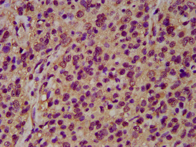
IHC image of diluted at 1:200 and staining in paraffin-embedded human glioma performed on a Leica BondTM system. After dewaxing and hydration, antigen retrieval was mediated by high pressure in a citrate buffer (pH 6.0). Section was blocked with 10% normal goat serum 30min at RT. Then primary antibody (1% BSA) was incubated at 4°C overnight. The primary is detected by a biotinylated secondary antibody and visualized using an HRP conjugated SP system.IF
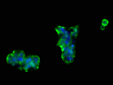
Immunofluorescence staining of 293 cells with at 1:66, counter-stained with DAPI. The cells were fixed in 4% formaldehyde, permeabilized using 0.2% Triton X-100 and blocked in 10% normal Goat Serum. The cells were then incubated with the antibody overnight at 4°C. The secondary antibody was Alexa Fluor 488-congugated AffiniPure Goat Anti-Rabbit IgG(H+L).| Product Name | NRCAM Antibody |
|---|---|
| Antibody Type | Primary Antibodies |
| Immunogen | Recombinant Human Centromere protein J protein (1082-1248AA) |
| Clonality | Polyclonal |
|---|---|
| Isotype | IgG |
| Host Species | Rabbit |
| Tested Applications | ELISAIFIHC |
| IHC:1:200-1:500 IF:1:50-1:200 |
|
| Species Reactivity | Human |
| Concentration | 1mg/ml |
| Purification | Protein G |
| Gene Symbol | NRCAM |
|---|---|
| Gene Synonyms | NEDNMS |
| Gene Full Name | neuronal cell adhesion molecule |
| Gene Summary | Cell adhesion molecules (CAMs) are members of the immunoglobulin superfamily. This gene encodes a neuronal cell adhesion molecule with multiple immunoglobulin-like C2-type domains and fibronectin type-III domains. This ankyrin-binding protein is involved in neuron-neuron adhesion and promotes directional signaling during axonal cone growth. This gene is also expressed in non-neural tissues and may play a general role in cell-cell communication via signaling from its intracellular domain to the actin cytoskeleton during directional cell migration. Allelic variants of this gene have been associated with autism and addiction vulnerability. Alternative splicing results in multiple transcript variants encoding different isoforms. [provided by RefSeq, Jul 2008] |
| Alternative Names | Bravo antibody hBravo antibody Neuronal cell adhesion molecule antibody Neuronal surface protein Bravo antibody Ng CAM related antibody Ng-CAM-related antibody NgCAM related cell adhesion molecule antibody NgCAM-related cell adhesion molecule antibody Nr CAM antibody Nr-CAM antibody Nrcam antibody NRCAM_HUMAN antibody |
| Cellular Localization | Cell membrane,Cell projection,Membrane,Secreted |
IHC
IHC image of diluted at 1:200 and staining in paraffin-embedded human glioma performed on a Leica BondTM system. After dewaxing and hydration, antigen retrieval was mediated by high pressure in a citrate buffer (pH 6.0). Section was blocked with 10% normal goat serum 30min at RT. Then primary antibody (1% BSA) was incubated at 4°C overnight. The primary is detected by a biotinylated secondary antibody and visualized using an HRP conjugated SP system.
IF
Immunofluorescence staining of 293 cells with at 1:66, counter-stained with DAPI. The cells were fixed in 4% formaldehyde, permeabilized using 0.2% Triton X-100 and blocked in 10% normal Goat Serum. The cells were then incubated with the antibody overnight at 4°C. The secondary antibody was Alexa Fluor 488-congugated AffiniPure Goat Anti-Rabbit IgG(H+L).| Application Notes | IHC:1:200-1:500 IF:1:50-1:200 |
|---|
| Form | Liquid |
|---|---|
| Storage Instructions | Upon receipt, store at -20°C or -80°C. Avoid repeated freeze. |
| Storage Buffer | Preservative: 0.03% Proclin 300,Constituents: 50% Glycerol, 0.01M PBS, pH 7.4 |


