WB
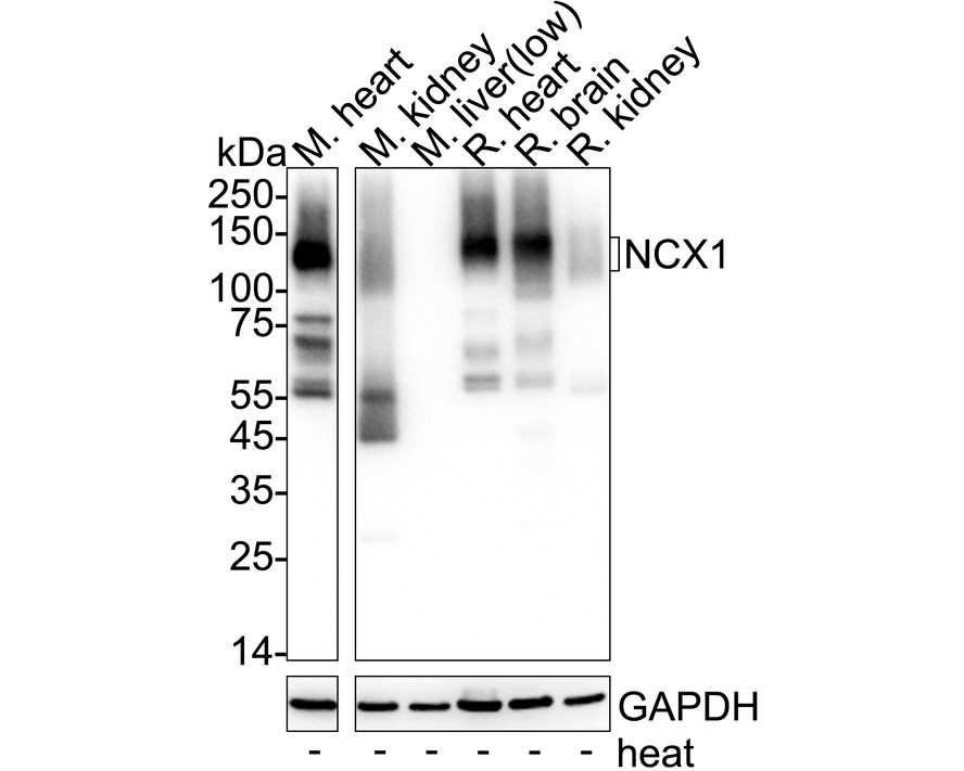
Western blot analysis of NCX1 on different lysates with Rabbit anti-NCX1 antibody at 1/5,000 dilution. Lane 1: Mouse heart tissue lysate Lane 2: Mouse kidney tissue lysate Lane 3: Mouse liver tissue lysate (low expression) Lane 4: Rat heart tissue lysate Lane 5: Rat brain tissue lysate Lane 6: Rat kidney tissue lysate Lysates/proteins at 20 µg/Lane. Predicted band size: 109 kDa Observed band size: 109 kDa Exposure time: 8 seconds; ECL: K1801; 4-20% SDS-PAGE gel. Proteins were transferred to a PVDF membrane and blocked with 5% NFDM/TBST for 1 hour at room temperature. The primary antibody at 1/5,000 dilution was used in primary antibody dilution at 4℃ overnight. Goat Anti-Rabbit IgG - HRP Secondary Antibody at 1/50,000 dilution was used for 1 hour at room temperature.IHC
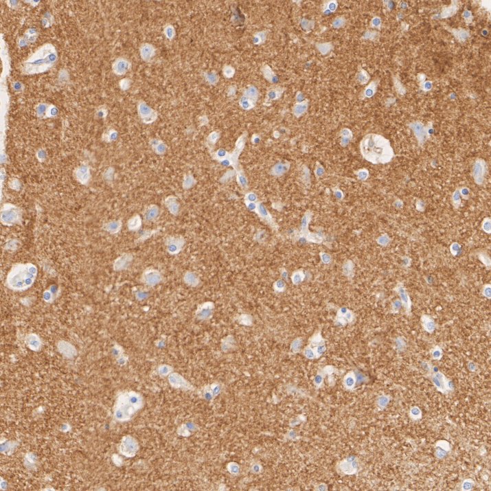
Immunohistochemical analysis of paraffin-embedded human brain tissue with Rabbit anti-NCX1 antibody at 1/1,000 dilution. The section was pre-treated using heat mediated antigen retrieval with Tris-EDTA buffer (pH 9.0) for 20 minutes. The tissues were blocked in 1% BSA for 20 minutes at room temperature, washed with ddH2O and PBS, and then probed with the primary antibody at 1/1,000 dilution for 1 hour at room temperature. The detection was performed using an HRP conjugated compact polymer system. DAB was used as the chromogen. Tissues were counterstained with hematoxylin and mounted with DPX.| Product Name | NCX1 Recombinant Rabbit Monoclonal Antibody |
|---|---|
| Antibody Type | Primary Antibodies |
| Immunogen | Recombinant protein within human NCX1 aa 253-800. |
| Clonality | Monoclonal |
|---|---|
| Isotype | IgG |
| Host Species | Rabbit |
| Tested Applications | IFIHCWB |
| WB:1:5,000 IHC:1:500 IF:1:200 |
|
| Species Reactivity | HumanMouseRat |
| Concentration | 1mg/ml |
| Purification | Protein A |
| Gene Symbol | SLC8A1 |
|---|---|
| Gene Synonyms | NCX1 |
| Gene Full Name | solute carrier family 8 member A1 |
| Gene Summary | In cardiac myocytes, Ca(2+) concentrations alternate between high levels during contraction and low levels during relaxation. The increase in Ca(2+) concentration during contraction is primarily due to release of Ca(2+) from intracellular stores. However, some Ca(2+) also enters the cell through the sarcolemma (plasma membrane). During relaxation, Ca(2+) is sequestered within the intracellular stores. To prevent overloading of intracellular stores, the Ca(2+) that entered across the sarcolemma must be extruded from the cell. The Na(+)-Ca(2+) exchanger is the primary mechanism by which the Ca(2+) is extruded from the cell during relaxation. In the heart, the exchanger may play a key role in digitalis action. The exchanger is the dominant mechanism in returning the cardiac myocyte to its resting state following excitation.[supplied by OMIM, Apr 2004] |
| Alternative Names | CNC antibody DKFZp779F0871 antibody MGC119581 antibody Na(+)/Ca(2+)-exchange protein 1 antibody Na+/Ca2+ exchange protein 1 antibody Na+/Ca2+ exchanger antibody NAC1_HUMAN antibody NCX 1 antibody NCX antibody NCX1 antibody CNC antibody DKFZp779F0871 antibody MGC119581 antibody Na(+)/Ca(2+)-exchange protein 1 antibody Na+/Ca2+ exchange protein 1 antibody Na+/Ca2+ exchanger antibody NAC1_HUMAN antibody NCX 1 antibody NCX antibody NCX1 antibody SLC8A1 antibody SLC8A1 protein antibody Sodium Calcium Exchanger antibody Sodium/calcium exchanger 1 antibody Solute carrier family 8 (sodium/calcium exchanger) member 1 antibody Solute carrier family 8 member 1 antibody |
| Molecular Weight(MW) | 109 kDa |
| Cellular Localization | Cell membrane,Membrane |
WB
Western blot analysis of NCX1 on different lysates with Rabbit anti-NCX1 antibody at 1/5,000 dilution. Lane 1: Mouse heart tissue lysate Lane 2: Mouse kidney tissue lysate Lane 3: Mouse liver tissue lysate (low expression) Lane 4: Rat heart tissue lysate Lane 5: Rat brain tissue lysate Lane 6: Rat kidney tissue lysate Lysates/proteins at 20 µg/Lane. Predicted band size: 109 kDa Observed band size: 109 kDa Exposure time: 8 seconds; ECL: K1801; 4-20% SDS-PAGE gel. Proteins were transferred to a PVDF membrane and blocked with 5% NFDM/TBST for 1 hour at room temperature. The primary antibody at 1/5,000 dilution was used in primary antibody dilution at 4℃ overnight. Goat Anti-Rabbit IgG - HRP Secondary Antibody at 1/50,000 dilution was used for 1 hour at room temperature.
IHC
Immunohistochemical analysis of paraffin-embedded human brain tissue with Rabbit anti-NCX1 antibody at 1/1,000 dilution. The section was pre-treated using heat mediated antigen retrieval with Tris-EDTA buffer (pH 9.0) for 20 minutes. The tissues were blocked in 1% BSA for 20 minutes at room temperature, washed with ddH2O and PBS, and then probed with the primary antibody at 1/1,000 dilution for 1 hour at room temperature. The detection was performed using an HRP conjugated compact polymer system. DAB was used as the chromogen. Tissues were counterstained with hematoxylin and mounted with DPX.| Application Notes | WB:1:5,000 IHC:1:500 IF:1:200 |
|---|
| Form | Liquid |
|---|---|
| Storage Instructions | Shipped at 4℃. Store at +4℃ short term (1-2 weeks). Store at -20℃ long term. |
| Storage Buffer | 1*PBS (pH7.4), 0.1% BSA, 40% Glycerol, 0.2% Proclean 950. |


