WB
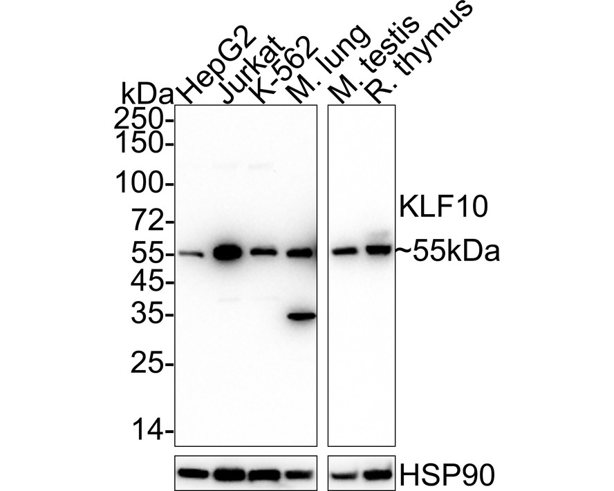
Western blot analysis of KLF10 on different lysates with Rabbit anti-KLF10 antibody at 1/1,000 dilution. Lane 1: HepG2 cell lysate (20 µg/Lane) Lane 2: Jurkat cell lysate (20 µg/Lane) Lane 3: K-562 cell lysate (20 µg/Lane) Lane 4: Mouse lung tissue lysate (40 µg/Lane) Lane 5: Mouse testis tissue lysate (40 µg/Lane) Lane 6: Rat thymus tissue lysate (40 µg/Lane) Predicted band size: 53 kDa Observed band size: 55 kDa Exposure time: Lane 1-4: 3 minutes; Lane 5-6: 30 seconds; ECL: K1801; 4-20% SDS-PAGE gel. Proteins were transferred to a PVDF membrane and blocked with 5% NFDM/TBST for 1 hour at room temperature. The primary antibody at 1/1,000 dilution was used in 5% NFDM/TBST at 4℃ overnight. Goat Anti-Rabbit IgG - HRP Secondary Antibody at 1/50,000 dilution was used for 1 hour at room temperature.| Product Name | KLF10 Recombinant Rabbit Monoclonal Antibody |
|---|---|
| Antibody Type | Primary Antibodies |
| Immunogen | Recombinant protein within Human KLF10 aa 1-100 / 480. |
| Clonality | Monoclonal |
|---|---|
| Isotype | IgG |
| Host Species | Rabbit |
| Tested Applications | WB |
| WB:1:1000 |
|
| Species Reactivity | HumanMouseRat |
| Concentration | 1mg/ml |
| Purification | Protein A |
| Gene Symbol | KLF10 |
|---|---|
| Gene Synonyms | EGRA TIEG TIEG1 EGR-alpha |
| Gene Full Name | KLF transcription factor 10 |
| Gene Summary | This gene encodes a member of a family of proteins that feature C2H2-type zinc finger domains. The encoded protein is a transcriptional repressor that acts as an effector of transforming growth factor beta signaling. Activity of this protein may inhibit the growth of cancers, particularly pancreatic cancer. Alternative splicing results in multiple transcript variants. [provided by RefSeq, Jun 2013] |
| Alternative Names | Early growth response alpha antibody Early growth response alpha like antibody EGR A antibody EGR alpha antibody EGR(A) antibody EGR-alpha antibody EGRA antibody Egral antibody EGRalpha antibody Gdnfif antibody Early growth response alpha antibody Early growth response alpha like antibody EGR A antibody EGR alpha antibody EGR(A) antibody EGR-alpha antibody EGRA antibody Egral antibody EGRalpha antibody Gdnfif antibody KLF 10 antibody KLF10 antibody KLF10_HUMAN antibody Krueppel like factor 10 antibody Krueppel like factor10 antibody Krueppel-like factor 10 antibody Kruppel like factor 10 antibody mGIF antibody TGF-beta- inducible early gene antibody TGFB inducible early growth response 1 antibody TGFB inducible early growth response antibody TGFB inducible early growth response protein 1 antibody TGFB-inducible early growth response protein 1 antibody TIEG 1 antibody TIEG antibody TIEG-1 antibody TIEG1 antibody Transforming growth factor beta inducible early growth response antibody Transforming growth factor beta inducible early growth response protein 1 antibody Transforming growth factor-beta-inducible early growth response protein 1 antibody Zinc finger transcription factor TIEG antibody |
| Molecular Weight(MW) | 53 kDa(Observed band size: 55 kDa) |
| Cellular Localization | Nucleus. |
WB
Western blot analysis of KLF10 on different lysates with Rabbit anti-KLF10 antibody at 1/1,000 dilution. Lane 1: HepG2 cell lysate (20 µg/Lane) Lane 2: Jurkat cell lysate (20 µg/Lane) Lane 3: K-562 cell lysate (20 µg/Lane) Lane 4: Mouse lung tissue lysate (40 µg/Lane) Lane 5: Mouse testis tissue lysate (40 µg/Lane) Lane 6: Rat thymus tissue lysate (40 µg/Lane) Predicted band size: 53 kDa Observed band size: 55 kDa Exposure time: Lane 1-4: 3 minutes; Lane 5-6: 30 seconds; ECL: K1801; 4-20% SDS-PAGE gel. Proteins were transferred to a PVDF membrane and blocked with 5% NFDM/TBST for 1 hour at room temperature. The primary antibody at 1/1,000 dilution was used in 5% NFDM/TBST at 4℃ overnight. Goat Anti-Rabbit IgG - HRP Secondary Antibody at 1/50,000 dilution was used for 1 hour at room temperature.| Application Notes | WB:1:1000 |
|---|
| Form | Liquid |
|---|---|
| Storage Instructions | Shipped at 4℃. Store at +4℃ short term (1-2 weeks). Store at -20℃ long term. |
| Storage Buffer | 1*TBS (pH7.4), 0.05% BSA, 40% Glycerol. Preservative: 0.05% Sodium Azide. |


