WB
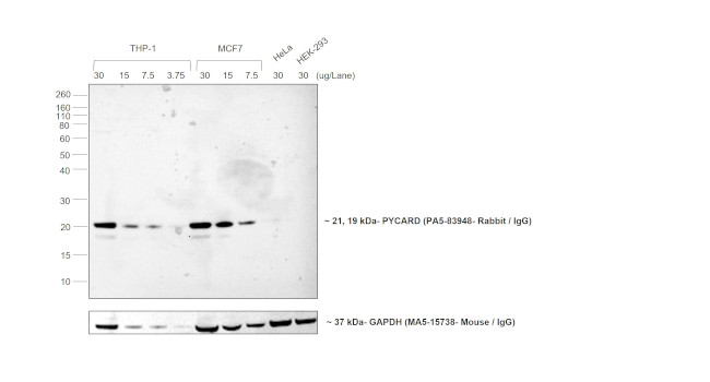
Chemiluminescent western blot was performed using Goat anti-Rabbit IgG (H+L) Cross-Adsorbed Secondary Antibody, Biotin-XX . Membrane enriched extracts of THP-1 (Lane 1, 2, 3, 4), MCF7 (Lane 5, 6, 7), HeLa (Lane 8) and HEK-293 (Lane 9) were electrophoresed usingNuPAGE™ 4-12% Bis-Tris Protein Gel. Resolved proteins were transferred onto anitrocellulose membrane byiBlot® 2 Dry BlottingSystem . The blot was probed with PYCARD Polyclonal Antibody. Secondary antibody was used for detection of PYCARD by chemiluminescence with SuperSignal™ West Pico PLUS Chemiluminescent Substrate using the iBright™ FL 1500. Poly-HRP Streptavidin (1:5000) was used to detect the biotin conjugated secondary antibody. After blocking the membrane, endogenous biotin was blocked using Streptavidin Protein ( 0.1 mg/mL (~1.7 μM)) for 15 minutes followed by addition of D- biotin (0.5 mg/mL (~2 mM)) for 1 hour, followed by primary antibody incubation. This ensures that the biotin in biological samples is masked and reduces the background. The Goat anti-Rabbit IgG (H+L) Cross-Adsorbed Secondary Antibody, Biotin-XX specifically detects the PYCARD rabbit primary antibody.ICC/IF
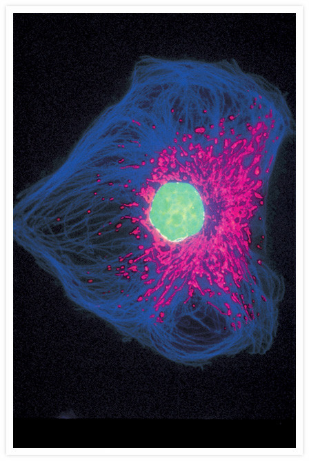
Bovine pulmonary artery endothelial cells incubated with the fixable, mitochondrion-selective MitoTracker® Red CMXRos . After staining, the cells were formaldehyde-fixed, acetone-permeabilized, treated with DNase-free RNase and counterstained using SYTOX® Green nucleic acid stain. Microtubules were labeled with a mouse monoclonal anti-β-tubulin antibody, biotin-XX goat anti-mouse IgG antibody and Cascade Blue® NeutrAvidin biotin-binding protein . This photograph was taken using multiple exposures through bandpass optical filters appropriate for Texas Red® dye, FITC and DAPI using a Nikon Labophot 2 microscope equipped with a Quadfluor epi-illumination system.IHC
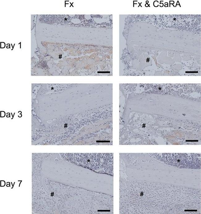
Immunostaining of IL-6 on day 1, 3 and 7 in tissue sections of control animals (Fx) and after administration of C5aRA showing the callus region in detail (fracture gap on the left, cortex in the middle, bone marrow on top (*) and periosteal callus (#) on the bottom of the image. Size of the scale bar = 100 µm (200-fold magnification) Image collected and cropped by CiteAb from the following publication (http://eurjmedres.biomedcentral.com/articles/10.1186/s40001-016-0236-7), licensed under a CC BY license.| Product Name | Goat anti-Rabbit IgG (H+L) Cross-Adsorbed Secondary Antibody, Biotin conjugated |
|---|---|
| Antibody Type | Secondary Antibodies |
| Clonality | Polyclonal |
|---|---|
| Isotype | IgG |
| Host Species | Rabbit |
| Tested Applications | ICC/IFIHCWB |
| WB:1:5000-1:10000 IHC: 1-10 µg/mL |
|
| Species Reactivity | Goat |
| Concentration | 2mg/ml |
| Purification | Affinity purified |
WB
Chemiluminescent western blot was performed using Goat anti-Rabbit IgG (H+L) Cross-Adsorbed Secondary Antibody, Biotin-XX . Membrane enriched extracts of THP-1 (Lane 1, 2, 3, 4), MCF7 (Lane 5, 6, 7), HeLa (Lane 8) and HEK-293 (Lane 9) were electrophoresed usingNuPAGE™ 4-12% Bis-Tris Protein Gel. Resolved proteins were transferred onto anitrocellulose membrane byiBlot® 2 Dry BlottingSystem . The blot was probed with PYCARD Polyclonal Antibody. Secondary antibody was used for detection of PYCARD by chemiluminescence with SuperSignal™ West Pico PLUS Chemiluminescent Substrate using the iBright™ FL 1500. Poly-HRP Streptavidin (1:5000) was used to detect the biotin conjugated secondary antibody. After blocking the membrane, endogenous biotin was blocked using Streptavidin Protein ( 0.1 mg/mL (~1.7 μM)) for 15 minutes followed by addition of D- biotin (0.5 mg/mL (~2 mM)) for 1 hour, followed by primary antibody incubation. This ensures that the biotin in biological samples is masked and reduces the background. The Goat anti-Rabbit IgG (H+L) Cross-Adsorbed Secondary Antibody, Biotin-XX specifically detects the PYCARD rabbit primary antibody.
ICC/IF
Bovine pulmonary artery endothelial cells incubated with the fixable, mitochondrion-selective MitoTracker® Red CMXRos . After staining, the cells were formaldehyde-fixed, acetone-permeabilized, treated with DNase-free RNase and counterstained using SYTOX® Green nucleic acid stain. Microtubules were labeled with a mouse monoclonal anti-β-tubulin antibody, biotin-XX goat anti-mouse IgG antibody and Cascade Blue® NeutrAvidin biotin-binding protein . This photograph was taken using multiple exposures through bandpass optical filters appropriate for Texas Red® dye, FITC and DAPI using a Nikon Labophot 2 microscope equipped with a Quadfluor epi-illumination system.
IHC
Immunostaining of IL-6 on day 1, 3 and 7 in tissue sections of control animals (Fx) and after administration of C5aRA showing the callus region in detail (fracture gap on the left, cortex in the middle, bone marrow on top (*) and periosteal callus (#) on the bottom of the image. Size of the scale bar = 100 µm (200-fold magnification) Image collected and cropped by CiteAb from the following publication (http://eurjmedres.biomedcentral.com/articles/10.1186/s40001-016-0236-7), licensed under a CC BY license.| Application Notes | WB:1:5000-1:10000 IHC: 1-10 µg/mL |
|---|
| Form | Liquid |
|---|---|
| Storage Instructions | Purified Goat IgG in PBS, pH 7.3, 0.01% sodium azide. |
| Storage Buffer | Store it at -20°C to -80°C for one year. It is recommended that the protein be aliquoted for optimal storage. Avoid repeated freeze-thaw cycles. |


