WB
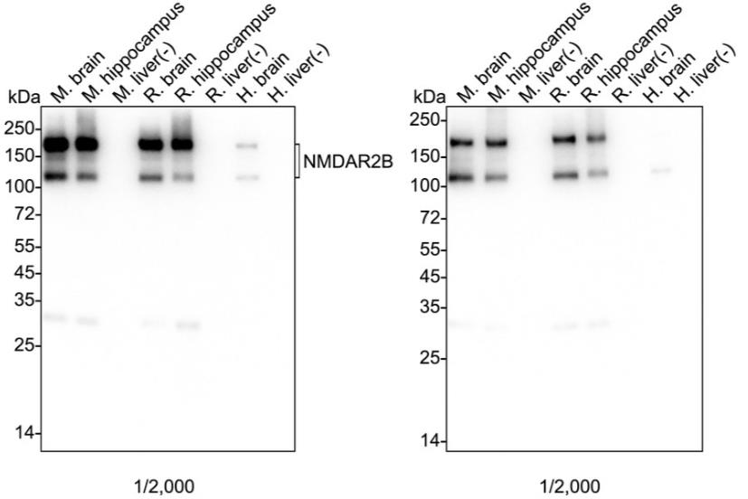
Western blot analysis of NMDAR2B on different lysates with Rabbit anti-NMDAR2B antibody at 1/2000 dilution and competitor's antibody at 1/2000 dilution. Lane 1: Mouse brain tissue lysate (no heat), Lane 2: Mouse hippocampus tissue lysate, Lane 3: Mouse liver tissue lysate (negative), Lane 4: Rat brain tissue lysate (no heat), Lane 5: Rat hippocampus tissue lysate, Lane 6: Rat liver tissue lysate (negative), Lane 7: Human brain tissue lysate, Lane 8: Human liver tissue lysate (negative), Notice: no heat means the lysate is not boiled. Lysates/proteins at 20 µg/Lane. Exposure time: 59 seconds; 4-20% SDS-PAGE gel. Proteins were transferred to a PVDF membrane and blocked with 5% NFDM/TBST for 1 hour at room temperature. The primary antibody at 1/2000 dilution and competitor's antibody at 1/2000 dilution were used in 5% NFDM/TBST at 4℃ overnight. Goat Anti-Rabbit IgG - HRP Secondary Antibody at 1/50,000 dilution was used for 1 hour at room temperature.IF-F
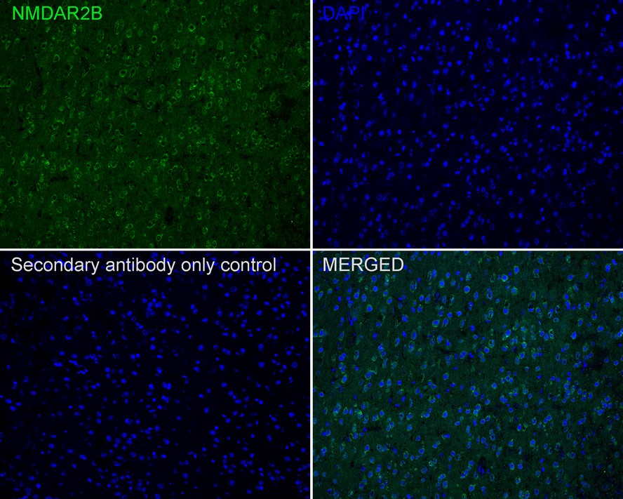
Immunofluorescence analysis of frozen mouse cerebellum tissue with Rabbit anti-NMDAR2B antibody at 1/500 dilution. The section was pre-treated using heat mediated antigen retrieval with sodium citrate buffer (pH 6.0) for about 2 minutes in microwave oven. The tissues were blocked in 10% negative goat serum for 1 hour at room temperature, washed with PBS, and then probed with the primary antibody (green) at 1/500 dilution overnight at 4 ℃, washed with PBS. Goat Anti-Rabbit IgG H&L (488) was used as the secondary antibody at 1/1000 dilution. Nuclei were counterstained with DAPI (blue).IHC
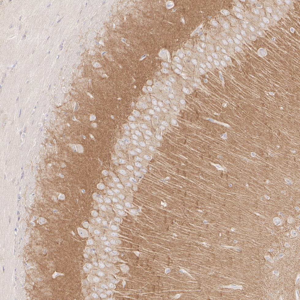
Immunohistochemical analysis of paraffin-embedded mouse hippocampus tissue with Rabbit anti-NMDAR2B antibody at 1/8000 dilution. The section was pre-treated using heat mediated antigen retrieval with Tris-EDTA buffer (pH 9.0) for 20 minutes. The tissues were blocked in 1% BSA for 20 minutes at room temperature, washed with ddH2O and PBS, and then probed with the primary antibody at 1/8000 dilution for 1 hour at room temperature. The detection was performed using an HRP conjugated compact polymer system. DAB was used as the chromogen. Tissues were counterstained with hematoxylin and mounted with DPX.IHC
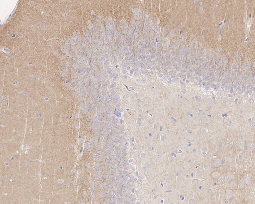
Immunohistochemical analysis of paraffin-embedded rat hippocampus tissue with Rabbit anti-NMDAR2B antibody at 1/2,000 dilution. The section was pre-treated using heat mediated antigen retrieval with Tris-EDTA buffer (pH 9.0) for 20 minutes. The tissues were blocked in 1% BSA for 20 minutes at room temperature, washed with ddH2O and PBS, and then probed with the primary antibody at 1/2000 dilution for 1 hour at room temperature. The detection was performed using an HRP conjugated compact polymer system. DAB was used as the chromogen. Tissues were counterstained with hematoxylin and mounted with DPX.| Product Name | NMDAR2B Recombinant Rabbit Monoclonal Antibody |
|---|---|
| Antibody Type | Primary Antibodies |
| Immunogen | Recombinant protein within human NMDAR2B aa 27-450 / 1,484. |
| Clonality | monoclonal |
|---|---|
| Isotype | IgG |
| Host Species | Rabbit |
| Tested Applications | IF-FIHCWB |
| WB:1:2000 IF-F:1:500 IHC:1:2000-1:8000 |
|
| Species Reactivity | HumanMouseRat |
| Concentration | 1mg/ml |
| Purification | Protein A |
| Gene Symbol | GRIN2B |
|---|---|
| Gene Synonyms | NR3 MRD6 NR2B hNR3 DEE27 EIEE27 GluN2B NMDAR2B |
| Gene Full Name | glutamate ionotropic receptor NMDA type subunit 2B |
| Gene Summary | This gene encodes a member of the N-methyl-D-aspartate (NMDA) receptor family within the ionotropic glutamate receptor superfamily. The encoded protein is a subunit of the NMDA receptor ion channel which acts as an agonist binding site for glutamate. The NMDA receptors mediate a slow calcium-permeable component of excitatory synaptic transmission in the central nervous system. The NMDA receptors are heterotetramers of seven genetically encoded, differentially expressed subunits including NR1 (GRIN1), NR2 (GRIN2A, GRIN2B, GRIN2C, or GRIN2D) and NR3 (GRIN3A or GRIN3B). The early expression of this gene in development suggests a role in brain development, circuit formation, synaptic plasticity, and cellular migration and differentiation. Naturally occurring mutations within this gene are associated with neurodevelopmental disorders including autism spectrum disorder, attention deficit hyperactivity disorder, epilepsy, and schizophrenia. [provided by RefSeq, Aug 2017] |
| Alternative Names | AW490526 antibody EIEE27 antibody Glutamate [NMDA] receptor subunit epsilon 2 antibody Glutamate [NMDA] receptor subunit epsilon-2 antibody Glutamate Receptor Ionotropic N Methyl D Aspartate 2B antibody Glutamate Receptor Ionotropic N Methyl D Aspartate subunit 2B antibody Glutamate receptor ionotropic NMDA2B antibody Glutamate receptor subunit epsilon 2 antibody Glutamate receptor ionotropic NMDA2B (epsilon 2) antibody GRIN 2B antibody AW490526 antibody EIEE27 antibody Glutamate [NMDA] receptor subunit epsilon 2 antibody Glutamate [NMDA] receptor subunit epsilon-2 antibody Glutamate Receptor Ionotropic N Methyl D Aspartate 2B antibody Glutamate Receptor Ionotropic N Methyl D Aspartate subunit 2B antibody Glutamate receptor ionotropic NMDA2B antibody Glutamate receptor subunit epsilon 2 antibody Glutamate receptor ionotropic NMDA2B (epsilon 2) antibody GRIN 2B antibody GRIN2B antibody hNR 3 antibody hNR3 antibody MGC142178 antibody MGC142180 antibody MRD6 antibody N methyl D asparate receptor channel subunit epsilon 2 antibody N methyl D aspartate receptor subtype 2B antibody N methyl D aspartate receptor subunit 2B antibody N methyl D aspartate receptor subunit 3 antibody N-methyl D-aspartate receptor subtype 2B antibody N-methyl-D-aspartate receptor subunit 3 antibody NMDA NR2B antibody NMDA R2B antibody NMDAR2B antibody NMDE2 antibody NMDE2_HUMAN antibody NME2 antibody NR2B antibody NR3 antibody |
| Molecular Weight(MW) | 166kDa(Observed band size:115/180kDa) |
| Cellular Localization | Cell membrane, Postsynaptic cell membrane, Late endosome, Lysosome, Cytoplasm, cytoskeleton. |
WB
Western blot analysis of NMDAR2B on different lysates with Rabbit anti-NMDAR2B antibody at 1/2000 dilution and competitor's antibody at 1/2000 dilution. Lane 1: Mouse brain tissue lysate (no heat), Lane 2: Mouse hippocampus tissue lysate, Lane 3: Mouse liver tissue lysate (negative), Lane 4: Rat brain tissue lysate (no heat), Lane 5: Rat hippocampus tissue lysate, Lane 6: Rat liver tissue lysate (negative), Lane 7: Human brain tissue lysate, Lane 8: Human liver tissue lysate (negative), Notice: no heat means the lysate is not boiled. Lysates/proteins at 20 µg/Lane. Exposure time: 59 seconds; 4-20% SDS-PAGE gel. Proteins were transferred to a PVDF membrane and blocked with 5% NFDM/TBST for 1 hour at room temperature. The primary antibody at 1/2000 dilution and competitor's antibody at 1/2000 dilution were used in 5% NFDM/TBST at 4℃ overnight. Goat Anti-Rabbit IgG - HRP Secondary Antibody at 1/50,000 dilution was used for 1 hour at room temperature.
IF-F
Immunofluorescence analysis of frozen mouse cerebellum tissue with Rabbit anti-NMDAR2B antibody at 1/500 dilution. The section was pre-treated using heat mediated antigen retrieval with sodium citrate buffer (pH 6.0) for about 2 minutes in microwave oven. The tissues were blocked in 10% negative goat serum for 1 hour at room temperature, washed with PBS, and then probed with the primary antibody (green) at 1/500 dilution overnight at 4 ℃, washed with PBS. Goat Anti-Rabbit IgG H&L (488) was used as the secondary antibody at 1/1000 dilution. Nuclei were counterstained with DAPI (blue).
IHC
Immunohistochemical analysis of paraffin-embedded mouse hippocampus tissue with Rabbit anti-NMDAR2B antibody at 1/8000 dilution. The section was pre-treated using heat mediated antigen retrieval with Tris-EDTA buffer (pH 9.0) for 20 minutes. The tissues were blocked in 1% BSA for 20 minutes at room temperature, washed with ddH2O and PBS, and then probed with the primary antibody at 1/8000 dilution for 1 hour at room temperature. The detection was performed using an HRP conjugated compact polymer system. DAB was used as the chromogen. Tissues were counterstained with hematoxylin and mounted with DPX.
IHC
Immunohistochemical analysis of paraffin-embedded rat hippocampus tissue with Rabbit anti-NMDAR2B antibody at 1/2,000 dilution. The section was pre-treated using heat mediated antigen retrieval with Tris-EDTA buffer (pH 9.0) for 20 minutes. The tissues were blocked in 1% BSA for 20 minutes at room temperature, washed with ddH2O and PBS, and then probed with the primary antibody at 1/2000 dilution for 1 hour at room temperature. The detection was performed using an HRP conjugated compact polymer system. DAB was used as the chromogen. Tissues were counterstained with hematoxylin and mounted with DPX.| Application Notes | WB:1:2000 IF-F:1:500 IHC:1:2000-1:8000 |
|---|
| Form | Liquid |
|---|---|
| Storage Instructions | Shipped at 4℃. Store at +4℃ short term (1-2 weeks). It is recommended to aliquot into single-use upon delivery. Store at -20℃ long term. |
| Storage Buffer | PBS (pH7.4), 0.1% BSA, 40% Glycerol. Preservative: 0.05% Sodium Azide. |


