WB
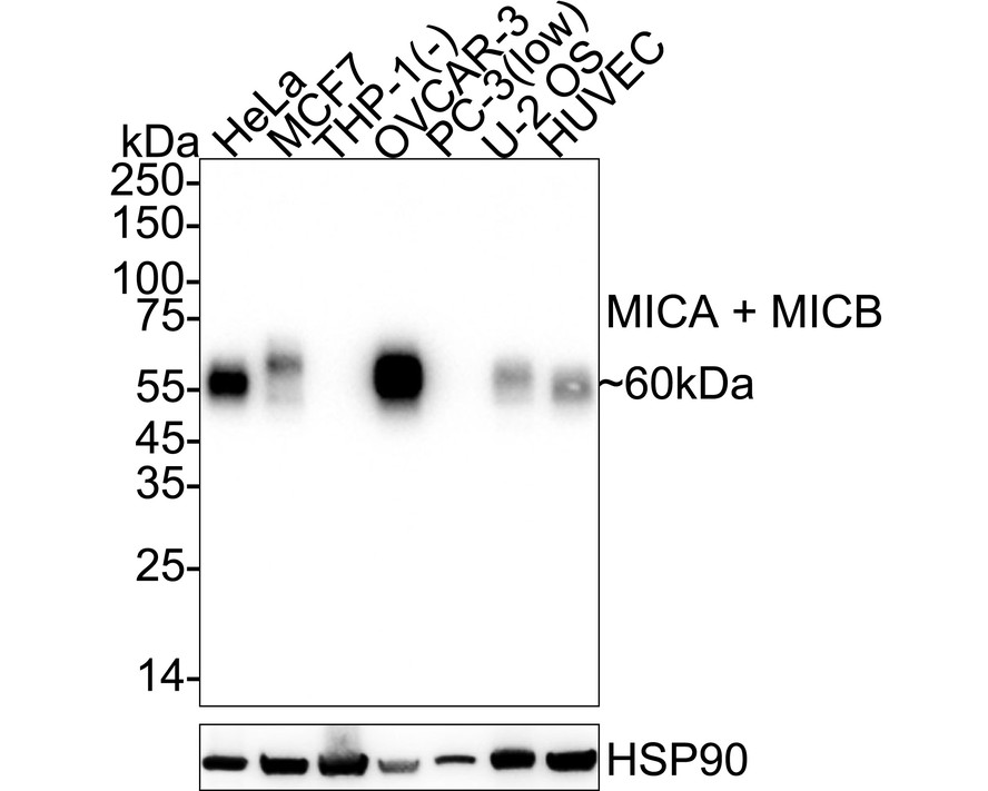
Western blot analysis of MICA + MICB on different lysates with Rabbit anti-MICA + MICB antibody at 1/5000 dilution. Lane 1: HeLa cell lysate (10 µg/Lane), Lane 2: MCF7 cell lysate (25 µg/Lane), Lane 3: THP-1 cell lysate (negative) (25 µg/Lane), Lane 4: NIH:OVCAR-3 cell lysate (25 µg/Lane), Lane 5: PC-3 cell lysate (low expression) (25 µg/Lane), Lane 6: U-2 OS cell lysate (25 µg/Lane), Lane 7: HUVEC cell lysate (25 µg/Lane), Exposure time: 20 seconds; 4-20% SDS-PAGE gel. Proteins were transferred to a PVDF membrane and blocked with 5% NFDM/TBST for 1 hour at room temperature. The primary antibody at 1/5,000 dilution was used in primary antibody dilution at 4℃ overnight. Goat Anti-Rabbit IgG - HRP Secondary Antibody at 1/50000 dilution was used for 1 hour at room temperature.WB
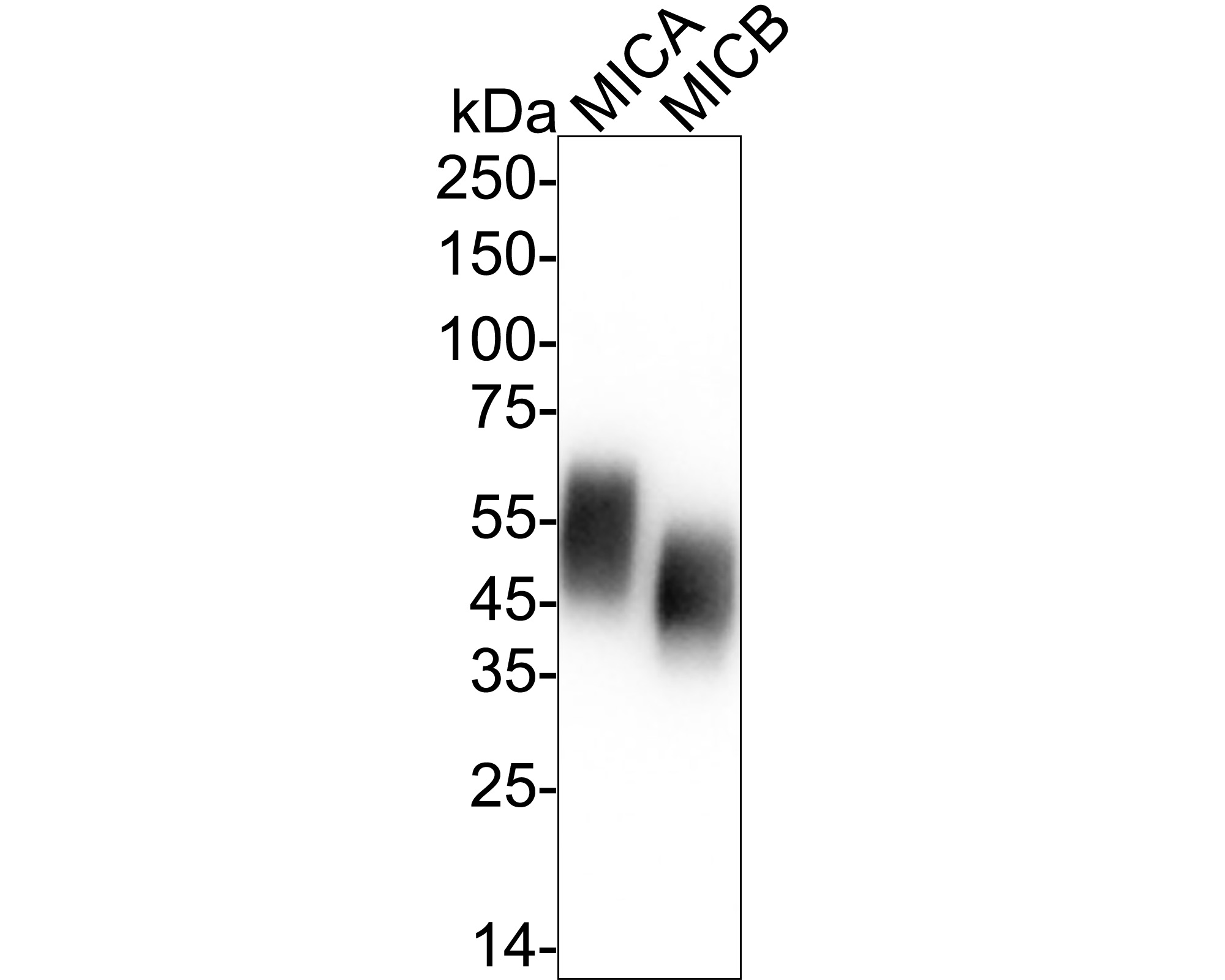
Western blot analysis of MICA + MICB on different lysates with Rabbit anti-MICA + MICB antibody at 1/2000 dilution. Lane 1: Human MICA recombinant protein, 20ng/Lane, Lane 2: Human MICB recombinant protein, 20ng/Lane, Exposure time: 2 seconds; 4-20% SDS-PAGE gel. Proteins were transferred to a PVDF membrane and blocked with 5% NFDM/TBST for 1 hour at room temperature. The primary antibody at 1/2000 dilution was used in 5% NFDM/TBST at 4℃ overnight. Goat Anti-Rabbit IgG - HRP Secondary Antibody at 1/50000 dilution was used for 1 hour at room temperature.FC
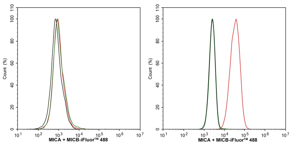
Flow cytometric analysis of THP-1 (left, negative) and HeLa (right, positive) cells labeling MICA + MICB. Cells were washed twice with cold PBS and resuspend. Then stained with the primary antibody (1/1000) (red) compared with Rabbit IgG Isotype Control (green). After incubation of the primary antibody at +4℃ for an hour, the cells were stained with a iFluor™ 488 conjugate-Goat anti-Rabbit IgG Secondary antibody at 1/1000 dilution for 30 minutes at +4℃. Unlabelled sample was used as a control (cells without incubation with primary antibody; black).IP
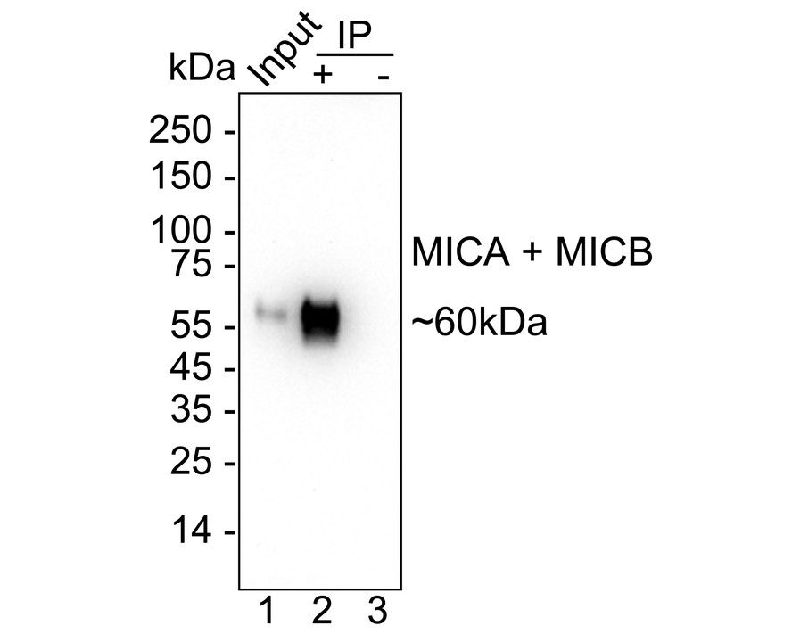
MICA + MICB was immunoprecipitated from 0.2 mg HeLa cell lysate with Rabbit anti-MICA + MICB antibody at 2 µg/10 µl beads. Western blot was performed from the immunoprecipitate using Rabbit anti-MICA + MICB antibody at 1/1,000 dilution. HRP Conjugated Anti-Rabbit IgG for IP Nano-secondary antibody at 1/5,000 dilution was used for 1 hour at room temperature. Lane 1: HeLa cell lysate (input), Lane 2:Rabbit anti-MICA + MICB antibody IP in HeLa cell lysate, Lane 3: Rabbit IgG instead of Rabbit anti-MICA + MICB antibody in HeLa cell lysate, Blocking/Dilution buffer: 5% NFDM/TBST Exposure time: 1 minute 7 seconds;| Product Name | MICA + MICB Recombinant Rabbit Monoclonal Antibody |
|---|---|
| Antibody Type | Primary Antibodies |
| Immunogen | Recombinant protein within human MICB aa 1-309. |
| Clonality | monoclonal |
|---|---|
| Isotype | IgG |
| Host Species | Rabbit |
| Tested Applications | FCIPWB |
| WB:1:5000 FC:1:1000 IP:1-2μg/sample |
|
| Species Reactivity | Human |
| Concentration | 1mg/ml |
| Purification | Protein A |
| Gene Symbol | MICB |
|---|---|
| Gene Synonyms | PERB11.2 |
| Gene Full Name | MHC class I polypeptide-related sequence B |
| Gene Summary | This gene encodes a heavily glycosylated protein which is a ligand for the NKG2D type II receptor. Binding of the ligand activates the cytolytic response of natural killer (NK) cells, CD8 alphabeta T cells, and gammadelta T cells which express the receptor. This protein is stress-induced and is similar to MHC class I molecules; however, it does not associate with beta-2-microglobulin or bind peptides. Alternative splicing results in multiple transcript variants. [provided by RefSeq, Jan 2014] |
| Alternative Names | HLA class I antigen antibody FLJ36918 antibody FLJ60820 antibody MGC111087 antibody MGC21250 antibody MHC class I chain related gene A protein antibody MHC class I chain related protein A antibody MHC class I chain related protein A HLA B HLA C antibody MHC class I polypeptide related sequence A antibody MHC class I polypeptide-related sequence A antibody HLA class I antigen antibody FLJ36918 antibody FLJ60820 antibody MGC111087 antibody MGC21250 antibody MHC class I chain related gene A protein antibody MHC class I chain related protein A antibody MHC class I chain related protein A HLA B HLA C antibody MHC class I polypeptide related sequence A antibody MHC class I polypeptide-related sequence A antibody MHC class I related protein antibody MIC A antibody MIC-A antibody micA antibody MICA_HUMAN antibody OTTHUMP00000029088 antibody OTTHUMP00000044528 antibody OTTHUMP00000165170 antibody OTTHUMP00000165172 antibody PERB11.1 antibody Stress inducible class I homolog antibody MHC class I chain related protein B antibody MHC class I chain related protein B2 antibody MHC class I like molecule PERB11.2 IMX antibody MHC class I mic B antigen antibody MHC class I polypeptide related sequence B antibody MHC class I polypeptide-related sequence B antibody MIC B antibody MIC-B antibody MICB antibody MICB_HUMAN antibody PERB11.2 antibody Stress inducible class I homolog antibody |
| Molecular Weight(MW) | 43kDa(Observed band size:60kDa) |
| Cellular Localization | Cell membrane, Cytoplasm; Cell membrane. |
WB
Western blot analysis of MICA + MICB on different lysates with Rabbit anti-MICA + MICB antibody at 1/5000 dilution. Lane 1: HeLa cell lysate (10 µg/Lane), Lane 2: MCF7 cell lysate (25 µg/Lane), Lane 3: THP-1 cell lysate (negative) (25 µg/Lane), Lane 4: NIH:OVCAR-3 cell lysate (25 µg/Lane), Lane 5: PC-3 cell lysate (low expression) (25 µg/Lane), Lane 6: U-2 OS cell lysate (25 µg/Lane), Lane 7: HUVEC cell lysate (25 µg/Lane), Exposure time: 20 seconds; 4-20% SDS-PAGE gel. Proteins were transferred to a PVDF membrane and blocked with 5% NFDM/TBST for 1 hour at room temperature. The primary antibody at 1/5,000 dilution was used in primary antibody dilution at 4℃ overnight. Goat Anti-Rabbit IgG - HRP Secondary Antibody at 1/50000 dilution was used for 1 hour at room temperature.
WB
Western blot analysis of MICA + MICB on different lysates with Rabbit anti-MICA + MICB antibody at 1/2000 dilution. Lane 1: Human MICA recombinant protein, 20ng/Lane, Lane 2: Human MICB recombinant protein, 20ng/Lane, Exposure time: 2 seconds; 4-20% SDS-PAGE gel. Proteins were transferred to a PVDF membrane and blocked with 5% NFDM/TBST for 1 hour at room temperature. The primary antibody at 1/2000 dilution was used in 5% NFDM/TBST at 4℃ overnight. Goat Anti-Rabbit IgG - HRP Secondary Antibody at 1/50000 dilution was used for 1 hour at room temperature.
FC
Flow cytometric analysis of THP-1 (left, negative) and HeLa (right, positive) cells labeling MICA + MICB. Cells were washed twice with cold PBS and resuspend. Then stained with the primary antibody (1/1000) (red) compared with Rabbit IgG Isotype Control (green). After incubation of the primary antibody at +4℃ for an hour, the cells were stained with a iFluor™ 488 conjugate-Goat anti-Rabbit IgG Secondary antibody at 1/1000 dilution for 30 minutes at +4℃. Unlabelled sample was used as a control (cells without incubation with primary antibody; black).
IP
MICA + MICB was immunoprecipitated from 0.2 mg HeLa cell lysate with Rabbit anti-MICA + MICB antibody at 2 µg/10 µl beads. Western blot was performed from the immunoprecipitate using Rabbit anti-MICA + MICB antibody at 1/1,000 dilution. HRP Conjugated Anti-Rabbit IgG for IP Nano-secondary antibody at 1/5,000 dilution was used for 1 hour at room temperature. Lane 1: HeLa cell lysate (input), Lane 2:Rabbit anti-MICA + MICB antibody IP in HeLa cell lysate, Lane 3: Rabbit IgG instead of Rabbit anti-MICA + MICB antibody in HeLa cell lysate, Blocking/Dilution buffer: 5% NFDM/TBST Exposure time: 1 minute 7 seconds;| Application Notes | WB:1:5000 FC:1:1000 IP:1-2μg/sample |
|---|
| Form | Liquid |
|---|---|
| Storage Instructions | Shipped at 4℃. Store at +4℃ short term (1-2 weeks). It is recommended to aliquot into single-use upon delivery. Store at -20℃ long term. |
| Storage Buffer | PBS (pH7.4), 0.1% BSA, 40% Glycerol. Preservative: 0.05% Sodium Azide. |


