WB
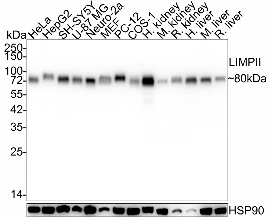
Western blot analysis of LIMPII on different lysates with Rabbit anti-LIMPII antibody at 1/2000 dilution. Lane 1: HeLa cell lysate (20 µg/Lane), Lane 2: HepG2 cell lysate (20 µg/Lane), Lane 3: SH-SY5Y cell lysate (20 µg/Lane), Lane 4: U-87 MG cell lysate (20 µg/Lane), Lane 5: Neuro-2a cell lysate (20 µg/Lane), Lane 6: MEF cell lysate (20 µg/Lane), Lane 7: PC-12 cell lysate (20 µg/Lane), Lane 8: COS-1 cell lysate (20 µg/Lane), Lane 9: Human kidney tissue lysate (40 µg/Lane), Lane 10: Mouse kidney tissue lysate (40 µg/Lane), Lane 11: Rat kidney tissue lysate (40 µg/Lane), Lane 12: Human liver tissue lysate (40 µg/Lane), Lane 13: Mouse liver tissue lysate (40 µg/Lane), Lane 14: Rat liver tissue lysate (40 µg/Lane), Exposure time: 5 seconds; 4-20% SDS-PAGE gel. Proteins were transferred to a PVDF membrane and blocked with 5% NFDM/TBST for 1 hour at room temperature. The primary antibody at 1/2000 dilution was used in 5% NFDM/TBST at 4℃ overnight. Goat Anti-Rabbit IgG - HRP Secondary Antibody at 1/50000 dilution was used for 1 hour at room temperature.ICC/IF
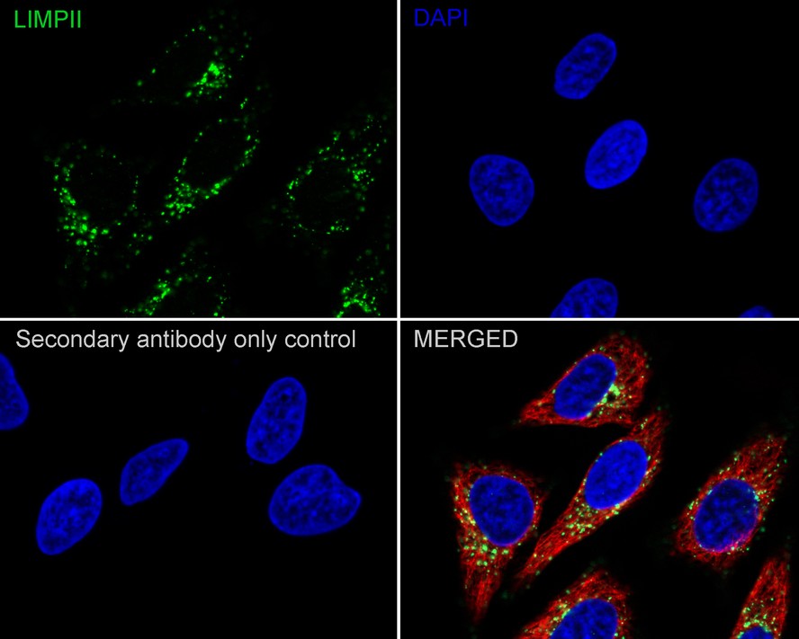
Immunocytochemistry analysis of HeLa cells labeling LIMPII with Rabbit anti-LIMPII antibody at 1/200 dilution. Cells were fixed in 100% precooled methanol for 5 minutes at room temperature, then blocked with 1% BSA in 10% negative goat serum for 1 hour at room temperature. Cells were then incubated with Rabbit anti-LIMPII antibody at 1/200 dilution in 1% BSA in PBST overnight at 4 ℃. Goat Anti-Rabbit IgG H&L (488) was used as the secondary antibody at 1/1,000 dilution. PBS instead of the primary antibody was used as the secondary antibody only control. Nuclear DNA was labelled in blue with DAPI. Beta tubulin (red) was stained at 1/100 dilution overnight at +4℃. Goat Anti-Mouse IgG H&L (594) was used as the secondary antibody at 1/1000 dilution.ICC/IF
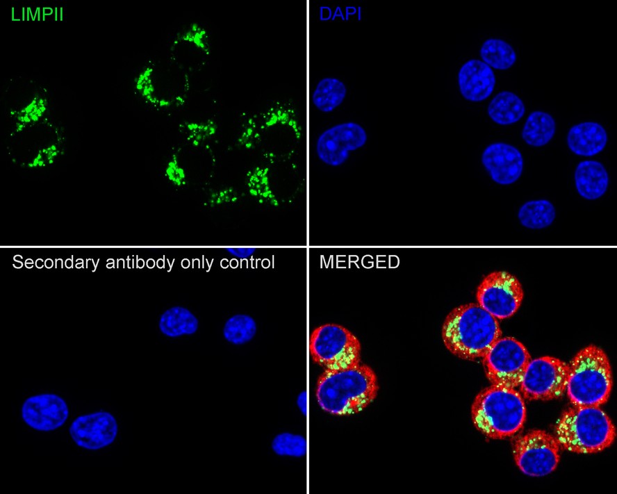
Immunocytochemistry analysis of Neuro-2a cells labeling LIMPII with Rabbit anti-LIMPII antibody at 1/200 dilution. Cells were fixed in 100% precooled methanol for 5 minutes at room temperature, then blocked with 1% BSA in 10% negative goat serum for 1 hour at room temperature. Cells were then incubated with Rabbit anti-LIMPII antibody at 1/200 dilution in 1% BSA in PBST overnight at 4 ℃. Goat Anti-Rabbit IgG H&L (488) was used as the secondary antibody at 1/1000 dilution. PBS instead of the primary antibody was used as the secondary antibody only control. Nuclear DNA was labelled in blue with DAPI. Beta tubulin (red) was stained at 1/100 dilution overnight at +4℃. Goat Anti-Mouse IgG H&L (594) was used as the secondary antibody at 1/1000 dilution.IHC
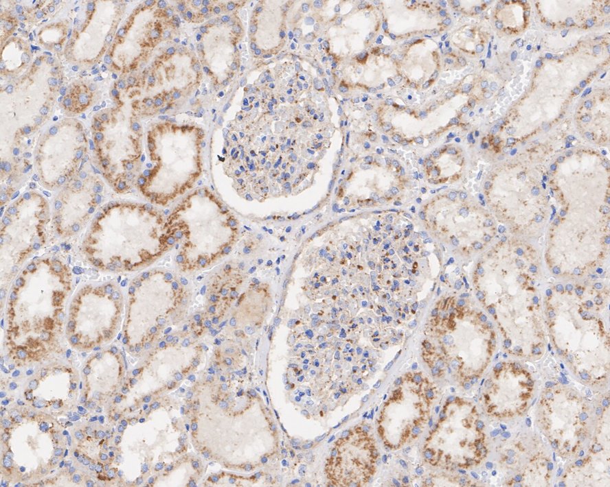
Immunohistochemical analysis of paraffin-embedded human kidney tissue with Rabbit anti-LIMPII antibody at 1/10000 dilution. The section was pre-treated using heat mediated antigen retrieval with Tris-EDTA buffer (pH 9.0) for 20 minutes. The tissues were blocked in 1% BSA for 20 minutes at room temperature, washed with ddH2O and PBS, and then probed with the primary antibody at 1/10000 dilution for 1 hour at room temperature. The detection was performed using an HRP conjugated compact polymer system. DAB was used as the chromogen. Tissues were counterstained with hematoxylin and mounted with DPX.IHC
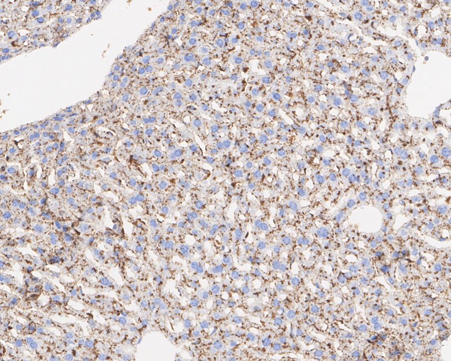
Immunohistochemical analysis of paraffin-embedded mouse liver tissue with Rabbit anti-LIMPII antibody at 1/10000 dilution. The section was pre-treated using heat mediated antigen retrieval with Tris-EDTA buffer (pH 9.0) for 20 minutes. The tissues were blocked in 1% BSA for 20 minutes at room temperature, washed with ddH2O and PBS, and then probed with the primary antibody at 1/10000 dilution for 1 hour at room temperature. The detection was performed using an HRP conjugated compact polymer system. DAB was used as the chromogen. Tissues were counterstained with hematoxylin and mounted with DPX.IHC
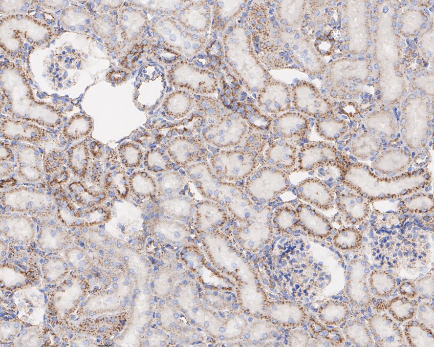
Immunohistochemical analysis of paraffin-embedded rat kidney tissue with Rabbit anti-LIMPII antibody at 1/10000 dilution. The section was pre-treated using heat mediated antigen retrieval with Tris-EDTA buffer (pH 9.0) for 20 minutes. The tissues were blocked in 1% BSA for 20 minutes at room temperature, washed with ddH2O and PBS, and then probed with the primary antibody at 1/10000 dilution for 1 hour at room temperature. The detection was performed using an HRP conjugated compact polymer system. DAB was used as the chromogen. Tissues were counterstained with hematoxylin and mounted with DPX.| Product Name | LIMPII Recombinant Rabbit Monoclonal Antibody |
|---|---|
| Antibody Type | Primary Antibodies |
| Product description |
|
| Immunogen | Recombinant protein within human LIMPII aa 5-459 / 478. |
| Clonality | monoclonal |
|---|---|
| Isotype | IgG |
| Host Species | Rabbit |
| Tested Applications | ICC/IFIHCWB |
| WB:1:2000-1:5000 ICC/IF:1:200 IHC:1:10000 |
|
| Species Reactivity | HumanMonkeyMouseRat |
| Concentration | 1mg/ml |
| Purification | Protein A |
| Gene Symbol | SCARB2 |
|---|---|
| Gene Synonyms | AMRF EPM4 LGP85 CD36L2 HLGP85 LIMP-2 LIMPII SR-BII |
| Gene Full Name | scavenger receptor class B member 2 |
| Gene Summary | The protein encoded by this gene is a type III glycoprotein that is located primarily in limiting membranes of lysosomes and endosomes. Earlier studies in mice and rat suggested that this protein may participate in membrane transportation and the reorganization of endosomal/lysosomal compartment. The protein deficiency in mice was reported to impair cell membrane transport processes and cause pelvic junction obstruction, deafness, and peripheral neuropathy. Further studies in human showed that this protein is a ubiquitously expressed protein and that it is involved in the pathogenesis of HFMD (hand, foot, and mouth disease) caused by enterovirus-71 and possibly by coxsackievirus A16. Mutations in this gene caused an autosomal recessive progressive myoclonic epilepsy-4 (EPM4), also known as action myoclonus-renal failure syndrome (AMRF). Alternatively spliced transcript variants encoding different isoforms have been found for this gene.[provided by RefSeq, Feb 2011] |
| Alternative Names | 85 kDa lysosomal membrane sialoglycoprotein antibody 85 kDa lysosomal sialoglycoprotein scavenger receptor class B member 2 antibody AMRF antibody CD36 antibody CD36 antigen (collagen type I receptor thrombospondin receptor)-like 2 (lysosomal integral membrane protein II) antibody CD36 antigen antibody CD36 antigen-like 2 antibody CD36L2 antibody EPM4 antibody HLGP85 antibody 85 kDa lysosomal membrane sialoglycoprotein antibody 85 kDa lysosomal sialoglycoprotein scavenger receptor class B member 2 antibody AMRF antibody CD36 antibody CD36 antigen (collagen type I receptor thrombospondin receptor)-like 2 (lysosomal integral membrane protein II) antibody CD36 antigen antibody CD36 antigen-like 2 antibody CD36L2 antibody EPM4 antibody HLGP85 antibody LGP85 antibody LIMP 2 antibody LIMP II antibody LIMP2 antibody LIMPII antibody Lysosomal integral membrane protein II antibody Lysosome membrane protein 2 antibody Lysosome membrane protein II antibody OTTHUMP00000160590 antibody OTTHUMP00000219176 antibody Scarb2 antibody Scavenger receptor class B member 2 antibody Scavenger receptor class B member 2 antibody SCRB2_HUMAN antibody SR BII antibody SRBII antibody |
| Molecular Weight(MW) | 54kDa(Observed band size: 80kDa) |
| Cellular Localization | Lysosome membrane. |
WB
Western blot analysis of LIMPII on different lysates with Rabbit anti-LIMPII antibody at 1/2000 dilution. Lane 1: HeLa cell lysate (20 µg/Lane), Lane 2: HepG2 cell lysate (20 µg/Lane), Lane 3: SH-SY5Y cell lysate (20 µg/Lane), Lane 4: U-87 MG cell lysate (20 µg/Lane), Lane 5: Neuro-2a cell lysate (20 µg/Lane), Lane 6: MEF cell lysate (20 µg/Lane), Lane 7: PC-12 cell lysate (20 µg/Lane), Lane 8: COS-1 cell lysate (20 µg/Lane), Lane 9: Human kidney tissue lysate (40 µg/Lane), Lane 10: Mouse kidney tissue lysate (40 µg/Lane), Lane 11: Rat kidney tissue lysate (40 µg/Lane), Lane 12: Human liver tissue lysate (40 µg/Lane), Lane 13: Mouse liver tissue lysate (40 µg/Lane), Lane 14: Rat liver tissue lysate (40 µg/Lane), Exposure time: 5 seconds; 4-20% SDS-PAGE gel. Proteins were transferred to a PVDF membrane and blocked with 5% NFDM/TBST for 1 hour at room temperature. The primary antibody at 1/2000 dilution was used in 5% NFDM/TBST at 4℃ overnight. Goat Anti-Rabbit IgG - HRP Secondary Antibody at 1/50000 dilution was used for 1 hour at room temperature.
ICC/IF
Immunocytochemistry analysis of HeLa cells labeling LIMPII with Rabbit anti-LIMPII antibody at 1/200 dilution. Cells were fixed in 100% precooled methanol for 5 minutes at room temperature, then blocked with 1% BSA in 10% negative goat serum for 1 hour at room temperature. Cells were then incubated with Rabbit anti-LIMPII antibody at 1/200 dilution in 1% BSA in PBST overnight at 4 ℃. Goat Anti-Rabbit IgG H&L (488) was used as the secondary antibody at 1/1,000 dilution. PBS instead of the primary antibody was used as the secondary antibody only control. Nuclear DNA was labelled in blue with DAPI. Beta tubulin (red) was stained at 1/100 dilution overnight at +4℃. Goat Anti-Mouse IgG H&L (594) was used as the secondary antibody at 1/1000 dilution.
ICC/IF
Immunocytochemistry analysis of Neuro-2a cells labeling LIMPII with Rabbit anti-LIMPII antibody at 1/200 dilution. Cells were fixed in 100% precooled methanol for 5 minutes at room temperature, then blocked with 1% BSA in 10% negative goat serum for 1 hour at room temperature. Cells were then incubated with Rabbit anti-LIMPII antibody at 1/200 dilution in 1% BSA in PBST overnight at 4 ℃. Goat Anti-Rabbit IgG H&L (488) was used as the secondary antibody at 1/1000 dilution. PBS instead of the primary antibody was used as the secondary antibody only control. Nuclear DNA was labelled in blue with DAPI. Beta tubulin (red) was stained at 1/100 dilution overnight at +4℃. Goat Anti-Mouse IgG H&L (594) was used as the secondary antibody at 1/1000 dilution.
IHC
Immunohistochemical analysis of paraffin-embedded human kidney tissue with Rabbit anti-LIMPII antibody at 1/10000 dilution. The section was pre-treated using heat mediated antigen retrieval with Tris-EDTA buffer (pH 9.0) for 20 minutes. The tissues were blocked in 1% BSA for 20 minutes at room temperature, washed with ddH2O and PBS, and then probed with the primary antibody at 1/10000 dilution for 1 hour at room temperature. The detection was performed using an HRP conjugated compact polymer system. DAB was used as the chromogen. Tissues were counterstained with hematoxylin and mounted with DPX.
IHC
Immunohistochemical analysis of paraffin-embedded mouse liver tissue with Rabbit anti-LIMPII antibody at 1/10000 dilution. The section was pre-treated using heat mediated antigen retrieval with Tris-EDTA buffer (pH 9.0) for 20 minutes. The tissues were blocked in 1% BSA for 20 minutes at room temperature, washed with ddH2O and PBS, and then probed with the primary antibody at 1/10000 dilution for 1 hour at room temperature. The detection was performed using an HRP conjugated compact polymer system. DAB was used as the chromogen. Tissues were counterstained with hematoxylin and mounted with DPX.
IHC
Immunohistochemical analysis of paraffin-embedded rat kidney tissue with Rabbit anti-LIMPII antibody at 1/10000 dilution. The section was pre-treated using heat mediated antigen retrieval with Tris-EDTA buffer (pH 9.0) for 20 minutes. The tissues were blocked in 1% BSA for 20 minutes at room temperature, washed with ddH2O and PBS, and then probed with the primary antibody at 1/10000 dilution for 1 hour at room temperature. The detection was performed using an HRP conjugated compact polymer system. DAB was used as the chromogen. Tissues were counterstained with hematoxylin and mounted with DPX.| Application Notes | WB:1:2000-1:5000 ICC/IF:1:200 IHC:1:10000 |
|---|
| Form | Liquid |
|---|---|
| Storage Instructions | Shipped at 4℃. Store at +4℃ short term (1-2 weeks). It is recommended to aliquot into single-use upon delivery. Store at -20℃ long term. |
| Storage Buffer | PBS (pH7.4), 0.1% BSA, 40% Glycerol. Preservative: 0.05% Sodium Azide. |


