WB
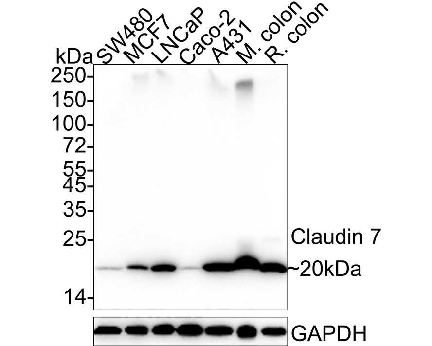
Western blot analysis of Claudin 7 on different lysates with Mouse anti-Claudin 7 antibody at 1/1000 dilution. Lane 1: SW480 cell lysate (20 µg/Lane), Lane 2: MCF7 cell lysate (20 µg/Lane), Lane 3: LNCaP cell lysate (20 µg/Lane), Lane 4: Caco-2 cell lysate (20 µg/Lane), Lane 5: A431 cell lysate (20 µg/Lane), Lane 6: Mouse colon tissue lysate (40 µg/Lane), Lane 7: Rat colon tissue lysate (40 µg/Lane), Exposure time: 1 minute; 4-20% SDS-PAGE gel. Proteins were transferred to a PVDF membrane and blocked with 5% NFDM/TBST for 1 hour at room temperature. The primary antibody at 1/1000 dilution was used in 5% NFDM/TBST at 4℃ overnight. Goat Anti-Mouse IgG - HRP Secondary Antibody at 1/50000 dilution was used for 1 hour at room temperature.ICC/IF
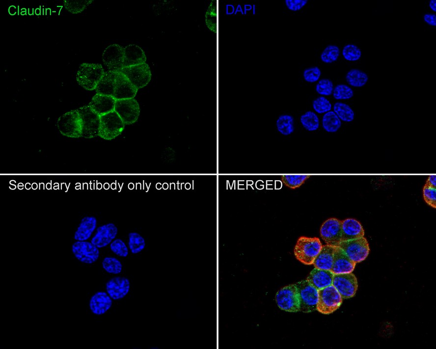
Immunocytochemistry analysis of PC-12 cells labeling Claudin 7 with Mouse anti-Claudin 7 antibody at 1/100 dilution. Cells were fixed in 4% paraformaldehyde for 20 minutes at room temperature, permeabilized with 0.1% Triton X-100 in PBS for 5 minutes at room temperature, then blocked with 1% BSA in 10% negative goat serum for 1 hour at room temperature. Cells were then incubated with Mouse anti-Claudin 7 antibody at 1/100 dilution in 1% BSA in PBST overnight at 4 ℃. Goat Anti-Mouse IgG H&L (488) was used as the secondary antibody at 1/1,000 dilution. PBS instead of the primary antibody was used as the secondary antibody only control. Nuclear DNA was labelled in blue with DAPI. beta Tubulin (red) was stained at 1/100 dilution overnight at +4℃. Goat Anti-Rabbit IgG H&L (594) were used as the secondary antibody at 1/1,000 dilution.IHC
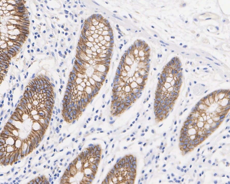
Immunohistochemical analysis of paraffin-embedded human colon tissue with Mouse anti-Claudin 7 antibody at 1/5000 dilution. The section was pre-treated using heat mediated antigen retrieval with Tris-EDTA buffer (pH 9.0) for 20 minutes. The tissues were blocked in 1% BSA for 20 minutes at room temperature, washed with ddH2O and PBS, and then probed with the primary antibody at 1/5000 dilution for 1 hour at room temperature. The detection was performed using an HRP conjugated compact polymer system. DAB was used as the chromogen. Tissues were counterstained with hematoxylin and mounted with DPX.IHC
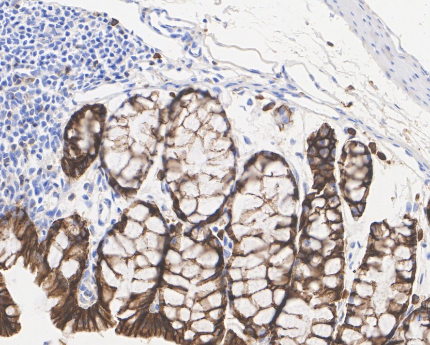
Immunohistochemical analysis of paraffin-embedded mouse colon tissue with Mouse anti-Claudin 7 antibody at 1/5000 dilution. The section was pre-treated using heat mediated antigen retrieval with Tris-EDTA buffer (pH 9.0) for 20 minutes. The tissues were blocked in 1% BSA for 20 minutes at room temperature, washed with ddH2O and PBS, and then probed with the primary antibody at 1/5000 dilution for 1 hour at room temperature. The detection was performed using an HRP conjugated compact polymer system. DAB was used as the chromogen. Tissues were counterstained with hematoxylin and mounted with DPX.IP
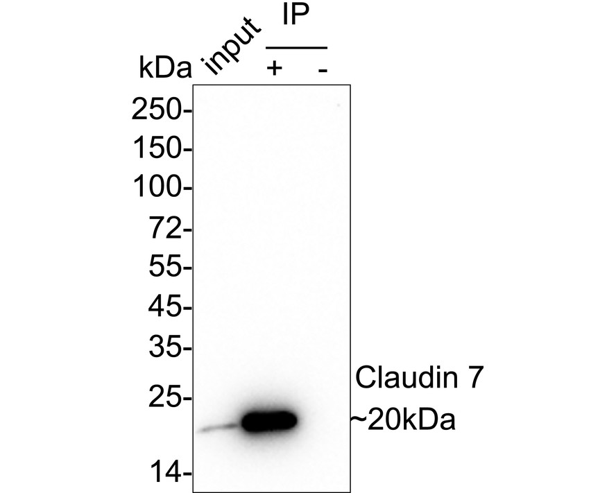
Claudin 7 was immunoprecipitated from 0.2 mg A431 cell lysate with Mouse anti-Claudin 7 antibody at 2 µg/10 µl beads. Western blot was performed from the immunoprecipitate using Mouse anti-Claudin 7 antibody at 1/2,000 dilution. Anti-Mouse IgG for IP Nano-secondary antibody at 1/5000 dilution was used for 1 hour at room temperature. Lane 1: A431 cell lysate (input) Lane 2:Mouse anti-Claudin 7 antibody IP in A431 cell lysate Lane 3: Mouse IgG instead of Mouse anti-Claudin 7 antibody in A431 cell lysate Blocking/Dilution buffer: 5% NFDM/TBST Exposure time: 2 minutes.| Product Name | Claudin 7 Recombinant Mouse Monoclonal Antibody |
|---|---|
| Antibody Type | Primary Antibodies |
| Product description |
|
| Immunogen | Synthetic peptide within Human Claudin 7 aa 162-211 / 211. |
| Clonality | Monoclonal |
|---|---|
| Isotype | IgG1 |
| Host Species | Mouse |
| Tested Applications | ICC/IFIHCIPWB |
| WB:1:1000 IHC:1:5000 ICC/IF:1:100 IP:1-2μg/sample |
|
| Species Reactivity | HumanMouseRat |
| Concentration | 1mg/ml |
| Purification | Protein A |
| Gene Symbol | CLDN7 |
|---|---|
| Gene Synonyms | CLDN-7 CEPTRL2 CPETRL2 Hs.84359 claudin-1 |
| Gene Full Name | claudin 7 |
| Gene Summary | This gene encodes a member of the claudin family. Claudins are integral membrane proteins and components of tight junction strands. Tight junction strands serve as a physical barrier to prevent solutes and water from passing freely through the paracellular space between epithelial or endothelial cell sheets, and also play critical roles in maintaining cell polarity and signal transductions. Differential expression of this gene has been observed in different types of malignancies, including breast cancer, ovarian cancer, hepatocellular carcinomas, urinary tumors, prostate cancer, lung cancer, head and neck cancers, thyroid carcinomas, etc.. Alternatively spliced transcript variants encoding different isoforms have been found.[provided by RefSeq, May 2010] |
| Alternative Names | CEPTR L2 antibody CEPTRL 2 antibody CEPTRL2 antibody Claudin 1 antibody Claudin 9 antibody Claudin-7 antibody Claudin1 antibody Claudin7 antibody Claudin9 antibody CLD7_HUMAN antibody CEPTR L2 antibody CEPTRL 2 antibody CEPTRL2 antibody Claudin 1 antibody Claudin 9 antibody Claudin-7 antibody Claudin1 antibody Claudin7 antibody Claudin9 antibody CLD7_HUMAN antibody CLDN 7 antibody CLDN-7 antibody CLDN7 antibody Clostridium perfringens enterotoxin receptor like 2 antibody CPETR L2 antibody CPETRL 2 antibody CPETRL2 antibody Hs.84359 antibody |
| Molecular Weight(MW) | 22kDa(Observed band size:20kDa) |
| Cellular Localization | Cell membrane, Basolateral cell membrane, Cell junction, tight junction. |
WB
Western blot analysis of Claudin 7 on different lysates with Mouse anti-Claudin 7 antibody at 1/1000 dilution. Lane 1: SW480 cell lysate (20 µg/Lane), Lane 2: MCF7 cell lysate (20 µg/Lane), Lane 3: LNCaP cell lysate (20 µg/Lane), Lane 4: Caco-2 cell lysate (20 µg/Lane), Lane 5: A431 cell lysate (20 µg/Lane), Lane 6: Mouse colon tissue lysate (40 µg/Lane), Lane 7: Rat colon tissue lysate (40 µg/Lane), Exposure time: 1 minute; 4-20% SDS-PAGE gel. Proteins were transferred to a PVDF membrane and blocked with 5% NFDM/TBST for 1 hour at room temperature. The primary antibody at 1/1000 dilution was used in 5% NFDM/TBST at 4℃ overnight. Goat Anti-Mouse IgG - HRP Secondary Antibody at 1/50000 dilution was used for 1 hour at room temperature.
ICC/IF
Immunocytochemistry analysis of PC-12 cells labeling Claudin 7 with Mouse anti-Claudin 7 antibody at 1/100 dilution. Cells were fixed in 4% paraformaldehyde for 20 minutes at room temperature, permeabilized with 0.1% Triton X-100 in PBS for 5 minutes at room temperature, then blocked with 1% BSA in 10% negative goat serum for 1 hour at room temperature. Cells were then incubated with Mouse anti-Claudin 7 antibody at 1/100 dilution in 1% BSA in PBST overnight at 4 ℃. Goat Anti-Mouse IgG H&L (488) was used as the secondary antibody at 1/1,000 dilution. PBS instead of the primary antibody was used as the secondary antibody only control. Nuclear DNA was labelled in blue with DAPI. beta Tubulin (red) was stained at 1/100 dilution overnight at +4℃. Goat Anti-Rabbit IgG H&L (594) were used as the secondary antibody at 1/1,000 dilution.
IHC
Immunohistochemical analysis of paraffin-embedded human colon tissue with Mouse anti-Claudin 7 antibody at 1/5000 dilution. The section was pre-treated using heat mediated antigen retrieval with Tris-EDTA buffer (pH 9.0) for 20 minutes. The tissues were blocked in 1% BSA for 20 minutes at room temperature, washed with ddH2O and PBS, and then probed with the primary antibody at 1/5000 dilution for 1 hour at room temperature. The detection was performed using an HRP conjugated compact polymer system. DAB was used as the chromogen. Tissues were counterstained with hematoxylin and mounted with DPX.
IHC
Immunohistochemical analysis of paraffin-embedded mouse colon tissue with Mouse anti-Claudin 7 antibody at 1/5000 dilution. The section was pre-treated using heat mediated antigen retrieval with Tris-EDTA buffer (pH 9.0) for 20 minutes. The tissues were blocked in 1% BSA for 20 minutes at room temperature, washed with ddH2O and PBS, and then probed with the primary antibody at 1/5000 dilution for 1 hour at room temperature. The detection was performed using an HRP conjugated compact polymer system. DAB was used as the chromogen. Tissues were counterstained with hematoxylin and mounted with DPX.
IP
Claudin 7 was immunoprecipitated from 0.2 mg A431 cell lysate with Mouse anti-Claudin 7 antibody at 2 µg/10 µl beads. Western blot was performed from the immunoprecipitate using Mouse anti-Claudin 7 antibody at 1/2,000 dilution. Anti-Mouse IgG for IP Nano-secondary antibody at 1/5000 dilution was used for 1 hour at room temperature. Lane 1: A431 cell lysate (input) Lane 2:Mouse anti-Claudin 7 antibody IP in A431 cell lysate Lane 3: Mouse IgG instead of Mouse anti-Claudin 7 antibody in A431 cell lysate Blocking/Dilution buffer: 5% NFDM/TBST Exposure time: 2 minutes.| Application Notes | WB:1:1000 IHC:1:5000 ICC/IF:1:100 IP:1-2μg/sample |
|---|
| Form | Liquid |
|---|---|
| Storage Instructions | Shipped at 4℃. Store at +4℃ short term (1-2 weeks). It is recommended to aliquot into single-use upon delivery. Store at -20℃ long term. |
| Storage Buffer | PBS (pH7.4), 0.1% BSA, 40% Glycerol. Preservative: 0.05% Sodium Azide. |


