WB
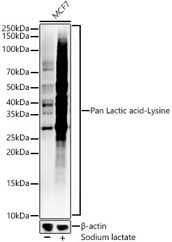
Western blot analysis of lysates from MCF7 cells, using Pan Lactic acid-Lysine Rabbit mAb at1:1000 dilution. MCF7 cells were treated with Sodium lactate(100mM) for 24h.Secondary antibody: HRP-conjugated Goat anti-Rabbit IgG (H+L) at 1:10000 dilution.Lysates/proteins: 25μg per lane.Blocking buffer: 3% nonfat dry milk in TBST.Detection: ECL Enhanced Kit.Exposure time: 180s.WB
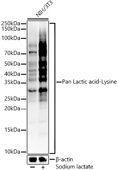
Western blot analysis of lysates from NIH/3T3 cells, using Pan Lactic acid-Lysine Rabbit mAb at1:1000 dilution. NIH/3T3 cells were treated with Sodium lactate(100mM) for 24h.Secondary antibody: HRP-conjugated Goat anti-Rabbit IgG (H+L) at 1:10000 dilution.Lysates/proteins: 25μg per lane.Blocking buffer: 3% nonfat dry milk in TBST.Detection: ECL Enhanced Kit.Exposure time: 180s.WB
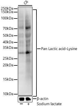
Western blot analysis of lysates from C6 cells, using Pan Lactic acid-Lysine Rabbit mAb at1:1000 dilution. C6 cells were treated with Sodium lactate(100mM) for 24h.Secondary antibody: HRP-conjugated Goat anti-Rabbit IgG (H+L) at 1:10000 dilution.Lysates/proteins: 25μg per lane.Blocking buffer: 3% nonfat dry milk in TBST.Detection: ECL Enhanced Kit .Exposure time: 180s.IP
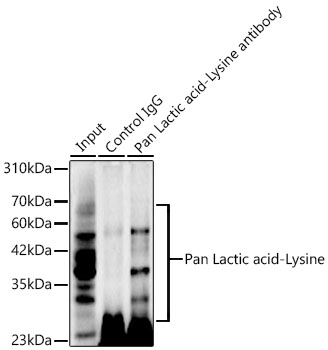
Immunoprecipitation of Pan Lactic acid-Lysine in 500 µg extracts from MCF7 cells trated by Sodiumlactate(200mM,24h) using 2 µg Pan Lactic acid-Lysine Rabbit mAb. Western blot analysis wasperformed using Pan Lactic acid-Lysine Rabbit mAb at 1:500 dilution.| Product Name | Pan Lactic acid-Lysine Rabbit mAb |
|---|---|
| Antibody Type | Primary Antibodies |
| Product description | Histone lysine lactation (Kla) is a newly discovered histone modification that regulates gene expression in macrophages. In M1 macrophages, lactic acid is derived from incompletely oxidized glucose and then produces lactyl-CoA, which is transferred via acetyltransferase p300 to the lysine tail of the histone. This modification is abundant in gene promoter regions that lack acetylation and are associated with gene expression activation. |
| Immunogen | Synthetic peptide. This information is considered to be commercially sensitive. |
| Clonality | Monoclonal |
|---|---|
| Isotype | IgG |
| Host Species | Rabbit |
| Tested Applications | IPWB |
| WB:1:500-1:1000 IP:0.5μg-4μg antibody for 400μg-600μg extracts of whole cells |
|
| Species Reactivity | HumanMouseOtherRat |
| Concentration | 1mg/ml |
| Purification | Affinity purified |
| Molecular Weight(MW) | Observed band size:10-250kDa |
|---|
WB
Western blot analysis of lysates from MCF7 cells, using Pan Lactic acid-Lysine Rabbit mAb at1:1000 dilution. MCF7 cells were treated with Sodium lactate(100mM) for 24h.Secondary antibody: HRP-conjugated Goat anti-Rabbit IgG (H+L) at 1:10000 dilution.Lysates/proteins: 25μg per lane.Blocking buffer: 3% nonfat dry milk in TBST.Detection: ECL Enhanced Kit.Exposure time: 180s.
WB
Western blot analysis of lysates from NIH/3T3 cells, using Pan Lactic acid-Lysine Rabbit mAb at1:1000 dilution. NIH/3T3 cells were treated with Sodium lactate(100mM) for 24h.Secondary antibody: HRP-conjugated Goat anti-Rabbit IgG (H+L) at 1:10000 dilution.Lysates/proteins: 25μg per lane.Blocking buffer: 3% nonfat dry milk in TBST.Detection: ECL Enhanced Kit.Exposure time: 180s.
WB
Western blot analysis of lysates from C6 cells, using Pan Lactic acid-Lysine Rabbit mAb at1:1000 dilution. C6 cells were treated with Sodium lactate(100mM) for 24h.Secondary antibody: HRP-conjugated Goat anti-Rabbit IgG (H+L) at 1:10000 dilution.Lysates/proteins: 25μg per lane.Blocking buffer: 3% nonfat dry milk in TBST.Detection: ECL Enhanced Kit .Exposure time: 180s.
IP
Immunoprecipitation of Pan Lactic acid-Lysine in 500 µg extracts from MCF7 cells trated by Sodiumlactate(200mM,24h) using 2 µg Pan Lactic acid-Lysine Rabbit mAb. Western blot analysis wasperformed using Pan Lactic acid-Lysine Rabbit mAb at 1:500 dilution.| Application Notes | WB:1:500-1:1000 IP:0.5μg-4μg antibody for 400μg-600μg extracts of whole cells |
|---|
| Form | Liquid |
|---|---|
| Storage Instructions | Store at -20℃. Avoid freeze / thaw cycles. |
| Storage Buffer | PBS with 0.09% Sodium azide, 0.05% BSA, 50% glycerol, pH7.3. |


