WB
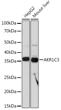
Western blot analysis of various lysates using AKR1C3 Rabbit mAb at 1:1000 dilution.Secondary antibody: HRP-conjugated Goat anti-Rabbit IgG (H+L) at 1:10000 dilution.Lysates/proteins: 25μg per lane.Blocking buffer: 3% nonfat dry milk in TBST.Detection: ECL Basic Kit.Exposure time: 10s.ICC/IF
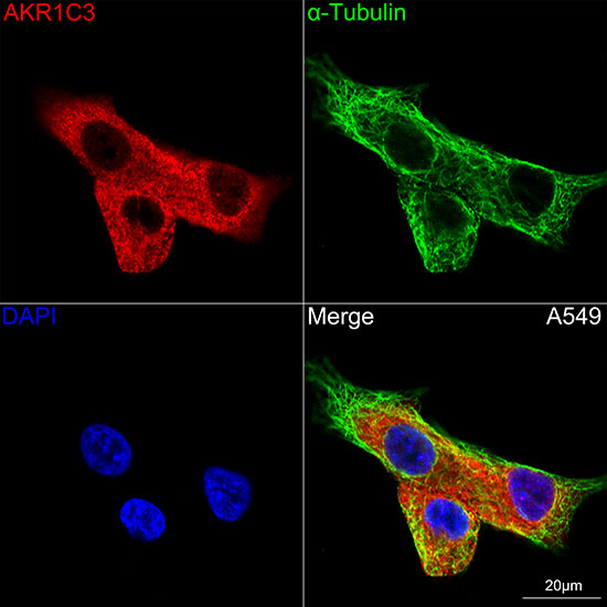
Confocal imaging of A549 cells using AKR1C3Rabbit mAb (dilution 1:100)(Red). Thecells were counterstained with α-TubulinMouse mAb (dilution 1:400) (Green).DAPI was used for nuclear staining (blue).Objective: 100x.| Product Name | AKR1C3 Rabbit mAb |
|---|---|
| Antibody Type | Primary Antibodies |
| Immunogen | Synthetic peptide. This information is considered to be commercially sensitive. |
| Clonality | Monoclonal |
|---|---|
| Isotype | IgG |
| Host Species | Rabbit |
| Tested Applications | ICC/IFWB |
| WB:1:1000-1:6000 ICC/IF:1:50-1:200 |
|
| Species Reactivity | HumanMouse |
| Concentration | 1mg/ml |
| Purification | Affinity purified |
| Gene Symbol | AKR1C3 |
|---|---|
| Gene Synonyms | DD3 DDX PGFS HAKRB HAKRe HA1753 HSD17B5 hluPGFS |
| Gene Full Name | aldo-keto reductase family 1 member C3 |
| Gene Summary | This gene encodes a member of the aldo/keto reductase superfamily, which consists of more than 40 known enzymes and proteins. These enzymes catalyze the conversion of aldehydes and ketones to their corresponding alcohols by utilizing NADH and/or NADPH as cofactors. The enzymes display overlapping but distinct substrate specificity. This enzyme catalyzes the reduction of prostaglandin (PG) D2, PGH2 and phenanthrenequinone (PQ), and the oxidation of 9alpha,11beta-PGF2 to PGD2. It may play an important role in the pathogenesis of allergic diseases such as asthma, and may also have a role in controlling cell growth and/or differentiation. This gene shares high sequence identity with three other gene members and is clustered with those three genes at chromosome 10p15-p14. Three transcript variants encoding different isoforms have been found for this gene. [provided by RefSeq, Dec 2011] |
| Alternative Names | DD3 DDX PGFS HAKRB HAKRe HA1753 HSD17B5 hluPGFS AKR1C3 |
| Molecular Weight(MW) | 37kDa(Observed band size:37kDa) |
| Cellular Localization | Cytoplasm. |
WB
Western blot analysis of various lysates using AKR1C3 Rabbit mAb at 1:1000 dilution.Secondary antibody: HRP-conjugated Goat anti-Rabbit IgG (H+L) at 1:10000 dilution.Lysates/proteins: 25μg per lane.Blocking buffer: 3% nonfat dry milk in TBST.Detection: ECL Basic Kit.Exposure time: 10s.
ICC/IF
Confocal imaging of A549 cells using AKR1C3Rabbit mAb (dilution 1:100)(Red). Thecells were counterstained with α-TubulinMouse mAb (dilution 1:400) (Green).DAPI was used for nuclear staining (blue).Objective: 100x.| Application Notes | WB:1:1000-1:6000 ICC/IF:1:50-1:200 |
|---|
| Form | Liquid |
|---|---|
| Storage Instructions | Store at -20℃. Avoid freeze / thaw cycles. |
| Storage Buffer | PBS containing 50% glycerol and 0.05% BSA, preserved with proclin300 or sodium azide (as specified on the Certificate of Analysis), pH 7.3. |


