WB
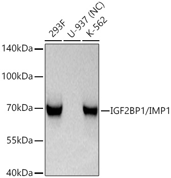
Western blot analysis of various lysates using IGF2BP1/IMP1 Rabbit mAb at 1:1000dilution incubated at room temperature for 1.5 hours.Secondary antibody: HRP-conjugated Goat anti-Rabbit IgG (H+L) at 1:10000 dilution.Lysates/proteins: 25 μg per lane.Blocking buffer: 3% nonfat dry milk in TBST.Detection: ECL Basic Kit.Negative control (NC): U-937Exposure time: 1s.WB
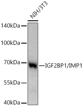
Western blot analysis of lysates from NIH/3T3 cells using IGF2BP1/IMP1 Rabbit mAb at 1:1000dilution incubated at room temperature for 1.5 hours.Secondary antibody: HRP-conjugated Goat anti-Rabbit IgG (H+L) at 1:10000 dilution.Lysates/proteins: 25 μg per lane.Blocking buffer: 3% nonfat dry milk in TBST.Detection: ECL Basic Kit.Exposure time: 30s.WB
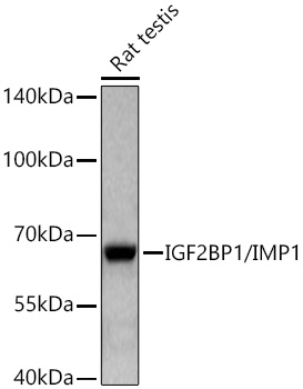
Western blot analysis of lysates from Rat testis using IGF2BP1/IMP1 Rabbit mAb at 1:1000dilution incubated at room temperature for 1.5 hours.Secondary antibody: HRP-conjugated Goat anti-Rabbit IgG (H+L) at 1:10000 dilution.Lysates/proteins: 25 μg per lane.Blocking buffer: 3% nonfat dry milk in TBST.Detection: ECL Basic Kit.Exposure time: 60s.ICC/IF
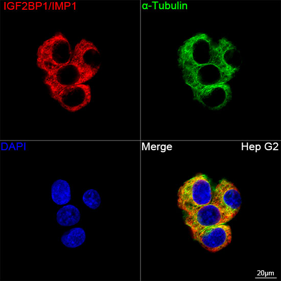
Confocal imaging of Hep G2 cells usingIGF2BP1/IMP1 Rabbit mAb (dilution1:200) followed by a further incubation withCy3 Goat Anti-Rabbit IgG (H+L) (dilution 1:500) (Red). The cells werecounterstained with α-Tubulin Mouse mAb(dilution 1:400) followed byincubation with ABflo® 488-conjugated GoatAnti-Mouse IgG (H+L) Ab (dilution1:500) (Green). DAPI was used for nuclearstaining (Blue). Objective: 100x.IHC
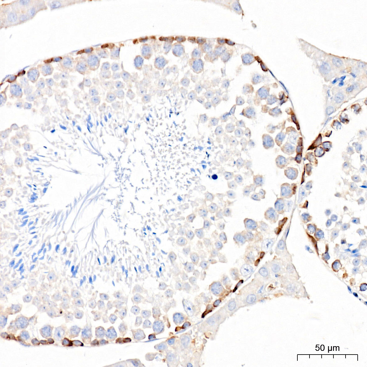
Immunohistochemistry analysis of paraffinembedded Mouse testis tissue usingIGF2BP1/IMP1 Rabbit mAb at adilution of 1:200 (40x lens). High pressureantigen retrieval performed with 0.01MCitrate buffer (pH 6.0) prior to IHC staining.IHC
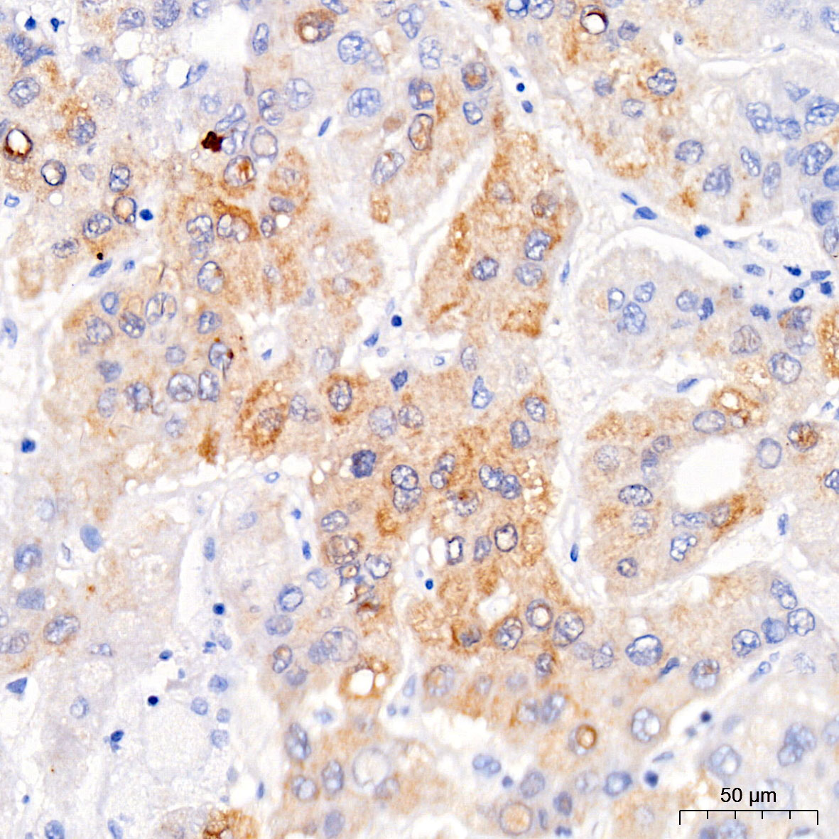
Immunohistochemistry analysis of paraffinembedded Human liver cancer tissue usingIGF2BP1/IMP1 Rabbit mAb at adilution of 1:200 (40x lens). High pressureantigen retrieval performed with 0.01MCitrate buffer (pH 6.0) prior to IHC staining.IHC
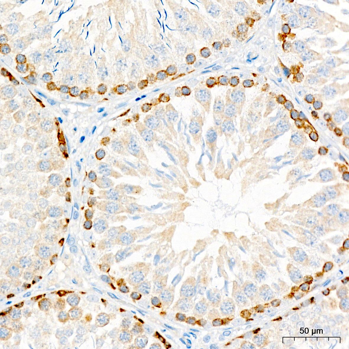
Immunohistochemistry analysis of paraffinembedded Rat testis tissue usingIGF2BP1/IMP1 Rabbit mAb at adilution of 1:200 (40x lens). High pressureantigen retrieval performed with 0.01MCitrate buffer (pH 6.0) prior to IHC staining.IP
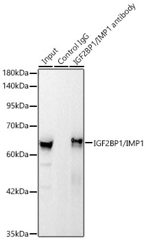
Immunoprecipitation of IGF2BP1/IMP1 from 500 µg extracts of 293F cells was performed using 2 µg ofIGF2BP1/IMP1 Rabbit mAb. Rabbit IgG isotype control was used to precipitate theControl IgG sample. IP samples were eluted with 1X non-reducing Laemmli Buffer. The Input lanerepresents 10% of the total input. Western blot analysis of immunoprecipitates was conducted usingIGF2BP1/IMP1 Rabbit mAb at a dilution of 1:1000.| Product Name | IGF2BP1/IMP1 Rabbit mAb |
|---|---|
| Antibody Type | Primary Antibodies |
| Immunogen | Synthetic peptide. This information is considered to be commercially sensitive. |
| Clonality | Monoclonal |
|---|---|
| Isotype | IgG |
| Host Species | Rabbit |
| Tested Applications | ICC/IFIHCIPWB |
| WB:1:1000-1:6000 IHC:1:200-1:800 ICC/IF:1:200-1:800 IP: 0.5μg-4μg antibody for 400μg-600μg extracts of whole cells |
|
| Species Reactivity | HumanMouseRat |
| Concentration | 1mg/ml |
| Purification | Affinity purified |
| Gene Symbol | IGF2BP1 |
|---|---|
| Gene Synonyms | IMP1 ZBP1 CRDBP IMP-1 CRD-BP VICKZ1 |
| Gene Full Name | insulin like growth factor 2 mRNA binding protein 1 |
| Gene Summary | This gene encodes a member of the insulin-like growth factor 2 mRNA-binding protein family. The protein encoded by this gene contains four K homology domains and two RNA recognition motifs. It functions by binding to the mRNAs of certain genes, including insulin-like growth factor 2, beta-actin and beta-transducin repeat-containing protein, and regulating their translation. Two transcript variants encoding different isoforms have been found for this gene. [provided by RefSeq, May 2009] |
| Alternative Names | IMP1 ZBP1 CRDBP IMP-1 CRD-BP VICKZ1 |
| Molecular Weight(MW) | 49/63kDa(Observed band size:64kDa) |
| Cellular Localization | Cell projection, Cytoplasm, Nucleus, axon, dendrite, dendritic spine, filopodium, growth cone, lamellipodium, perinuclear region, CRD-mediated mRNA stability complex, cytoplasm, cytoplasmic stress granule, cytosol, nucleoplasm, nucleus, P-body, perinuclear region of cytoplasm. |
WB
Western blot analysis of various lysates using IGF2BP1/IMP1 Rabbit mAb at 1:1000dilution incubated at room temperature for 1.5 hours.Secondary antibody: HRP-conjugated Goat anti-Rabbit IgG (H+L) at 1:10000 dilution.Lysates/proteins: 25 μg per lane.Blocking buffer: 3% nonfat dry milk in TBST.Detection: ECL Basic Kit.Negative control (NC): U-937Exposure time: 1s.
WB
Western blot analysis of lysates from NIH/3T3 cells using IGF2BP1/IMP1 Rabbit mAb at 1:1000dilution incubated at room temperature for 1.5 hours.Secondary antibody: HRP-conjugated Goat anti-Rabbit IgG (H+L) at 1:10000 dilution.Lysates/proteins: 25 μg per lane.Blocking buffer: 3% nonfat dry milk in TBST.Detection: ECL Basic Kit.Exposure time: 30s.
WB
Western blot analysis of lysates from Rat testis using IGF2BP1/IMP1 Rabbit mAb at 1:1000dilution incubated at room temperature for 1.5 hours.Secondary antibody: HRP-conjugated Goat anti-Rabbit IgG (H+L) at 1:10000 dilution.Lysates/proteins: 25 μg per lane.Blocking buffer: 3% nonfat dry milk in TBST.Detection: ECL Basic Kit.Exposure time: 60s.
ICC/IF
Confocal imaging of Hep G2 cells usingIGF2BP1/IMP1 Rabbit mAb (dilution1:200) followed by a further incubation withCy3 Goat Anti-Rabbit IgG (H+L) (dilution 1:500) (Red). The cells werecounterstained with α-Tubulin Mouse mAb(dilution 1:400) followed byincubation with ABflo® 488-conjugated GoatAnti-Mouse IgG (H+L) Ab (dilution1:500) (Green). DAPI was used for nuclearstaining (Blue). Objective: 100x.
IHC
Immunohistochemistry analysis of paraffinembedded Mouse testis tissue usingIGF2BP1/IMP1 Rabbit mAb at adilution of 1:200 (40x lens). High pressureantigen retrieval performed with 0.01MCitrate buffer (pH 6.0) prior to IHC staining.
IHC
Immunohistochemistry analysis of paraffinembedded Human liver cancer tissue usingIGF2BP1/IMP1 Rabbit mAb at adilution of 1:200 (40x lens). High pressureantigen retrieval performed with 0.01MCitrate buffer (pH 6.0) prior to IHC staining.
IHC
Immunohistochemistry analysis of paraffinembedded Rat testis tissue usingIGF2BP1/IMP1 Rabbit mAb at adilution of 1:200 (40x lens). High pressureantigen retrieval performed with 0.01MCitrate buffer (pH 6.0) prior to IHC staining.
IP
Immunoprecipitation of IGF2BP1/IMP1 from 500 µg extracts of 293F cells was performed using 2 µg ofIGF2BP1/IMP1 Rabbit mAb. Rabbit IgG isotype control was used to precipitate theControl IgG sample. IP samples were eluted with 1X non-reducing Laemmli Buffer. The Input lanerepresents 10% of the total input. Western blot analysis of immunoprecipitates was conducted usingIGF2BP1/IMP1 Rabbit mAb at a dilution of 1:1000.| Application Notes | WB:1:1000-1:6000 IHC:1:200-1:800 ICC/IF:1:200-1:800 IP: 0.5μg-4μg antibody for 400μg-600μg extracts of whole cells |
|---|
| Form | Liquid |
|---|---|
| Storage Instructions | Store at -20℃. Avoid freeze / thaw cycles. |
| Storage Buffer | PBS with 0.09% Sodium azide, 0.05% BSA, 50% glycerol, pH7.3. |


