WB
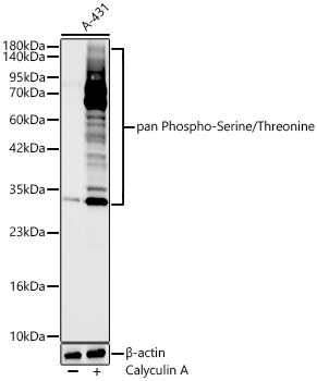
Western blot analysis of lysates from A-431 cells using pan Phospho-Serine/Threonine Rabbit mAb at 1:1000 dilution. A-431 cells were treated with Calyculin A (200 nM) at 37℃ for 30 minutesafter serum-starvation overnight.Secondary antibody: HRP-conjugated Goat anti-Rabbit IgG (H+L) at 1:10000 dilution.Lysates/proteins: 25 μg per lane.Blocking buffer: 3% nonfat dry milk in TBST.Detection: ECL Basic Kit.Exposure time: 90s.IHC
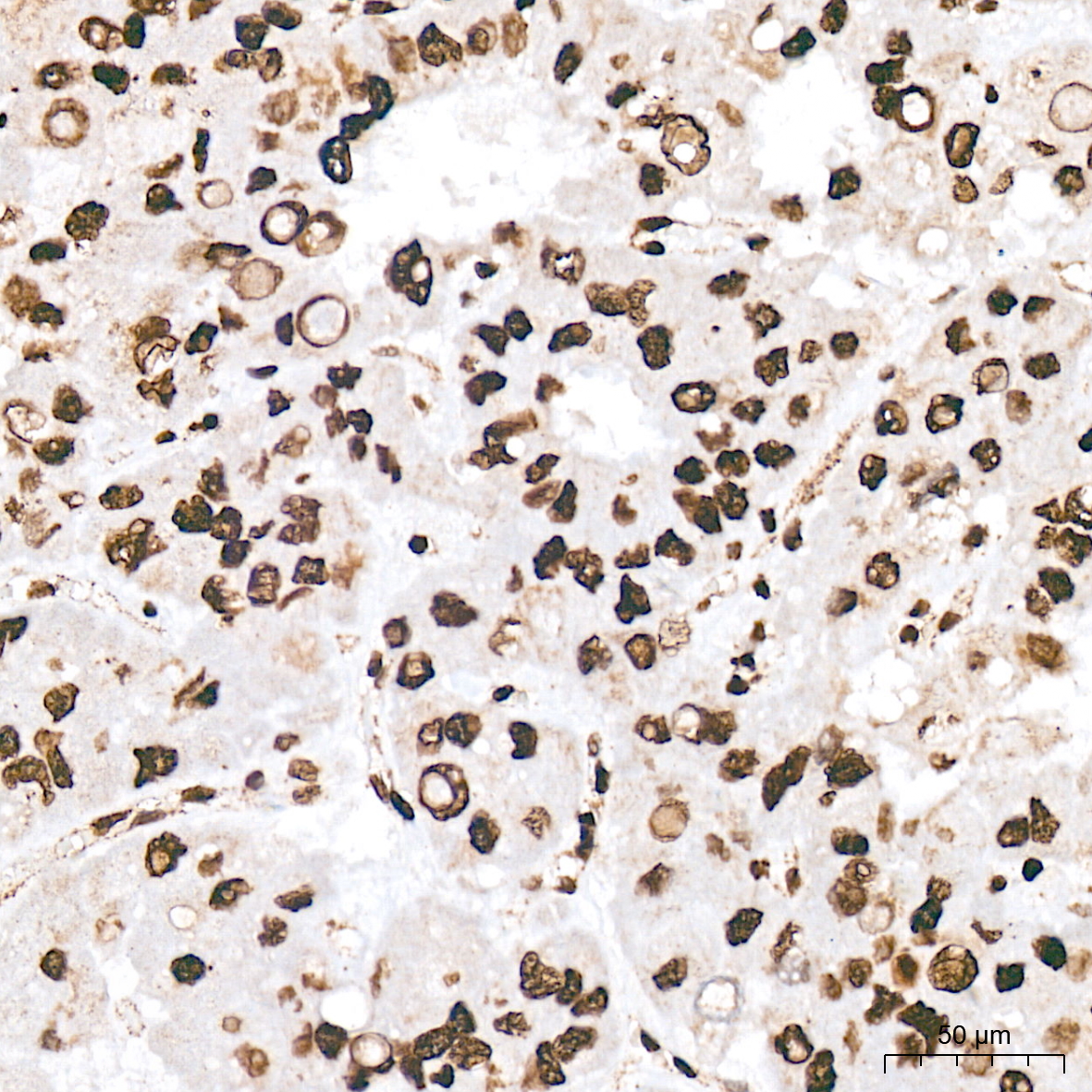
Immunohistochemistry analysis of paraffinembedded Human liver tissue using panPhospho-Serine/Threonine RabbitmAb at a dilution of 1:200 (40xlens). High pressure antigen retrieval wasperformed with 0.01 M citrate buffer (pH 6.0)prior to IHC staining.IHC
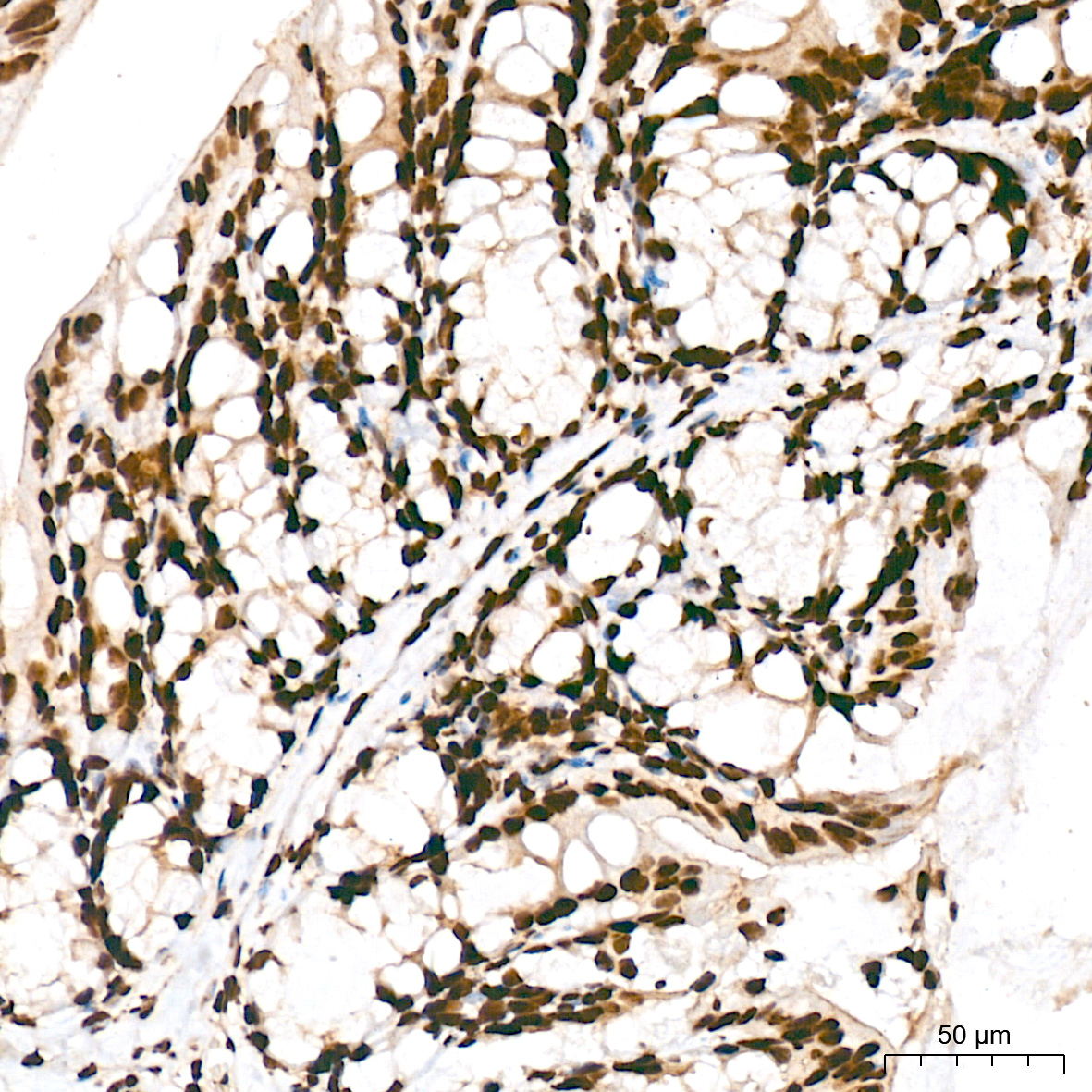
Immunohistochemistry analysis of paraffinembedded Mouse colon tissue using panPhospho-Serine/Threonine RabbitmAb at a dilution of 1:200 (40xlens). High pressure antigen retrieval wasperformed with 0.01 M citrate buffer (pH 6.0)prior to IHC staining.IHC
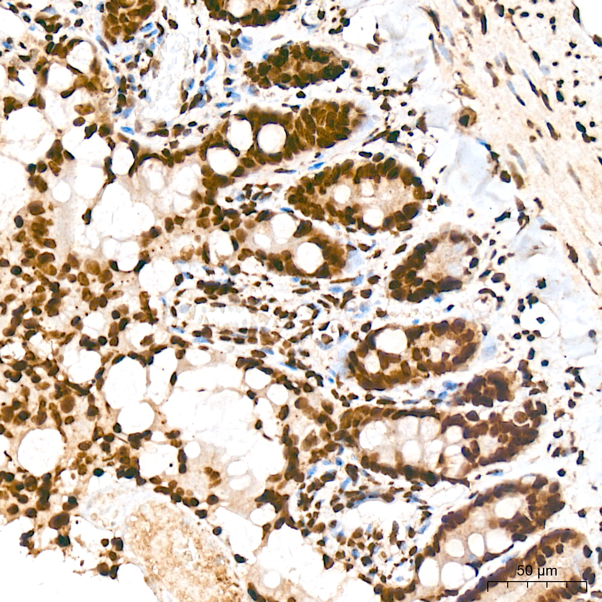
Immunohistochemistry analysis of paraffinembedded Rat colon tissue using panPhospho-Serine/Threonine RabbitmAb at a dilution of 1:200 (40xlens). High pressure antigen retrieval wasperformed with 0.01 M citrate buffer (pH 6.0)prior to IHC staining.IP
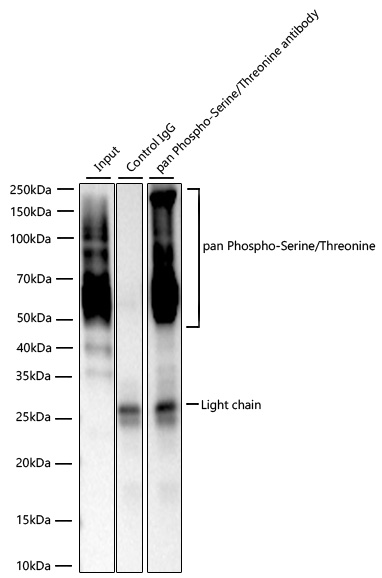
Immunoprecipitation of pan Phospho-Serine/Threonine from 300 µg extracts of A-431 cells treated withcalyculin A (200nM,30min) was performed using 2 µg of pan Phospho-Serine/Threonine Rabbit mAb. Rabbit Control IgG was used to precipitate the Control IgG sample. IP samples wereeluted with 1x reducing Laemmli Buffer. The Input lane represents 10% of the total input. Western blotanalysis of immunoprecipitates was conducted using pan Phospho-Serine/Threonine Rabbit mAb at a dilution of 1:3000.| Product Name | pan Phospho-Serine/Threonine Rabbit mAb |
|---|---|
| Antibody Type | Primary Antibodies |
| Product description | Protein phosphorylation is one of the main key regulatory mechanisms by which extracellular signals are conveyed. Global alteration of signal transduction by inhibition of serine/threonine dephosphorylation has recently been shown to markedly potentiate cancer cell killing by the DNA-methylating drug, temozolomide. |
| Immunogen | Synthetic peptide. This information is considered to be commercially sensitive. |
| Clonality | Monoclonal |
|---|---|
| Isotype | IgG |
| Host Species | Rabbit |
| Tested Applications | IHCIPWB |
| WB:1:1000-1:4000 IHC:1:50-1:200 IP:2μg-4μg antibody for 200μg-400μg extracts of whole cells |
|
| Species Reactivity | HumanMouseOtherRat |
| Concentration | 1mg/ml |
| Purification | Affinity purified |
| Molecular Weight(MW) | >10kDa |
|---|
WB
Western blot analysis of lysates from A-431 cells using pan Phospho-Serine/Threonine Rabbit mAb at 1:1000 dilution. A-431 cells were treated with Calyculin A (200 nM) at 37℃ for 30 minutesafter serum-starvation overnight.Secondary antibody: HRP-conjugated Goat anti-Rabbit IgG (H+L) at 1:10000 dilution.Lysates/proteins: 25 μg per lane.Blocking buffer: 3% nonfat dry milk in TBST.Detection: ECL Basic Kit.Exposure time: 90s.
IHC
Immunohistochemistry analysis of paraffinembedded Human liver tissue using panPhospho-Serine/Threonine RabbitmAb at a dilution of 1:200 (40xlens). High pressure antigen retrieval wasperformed with 0.01 M citrate buffer (pH 6.0)prior to IHC staining.
IHC
Immunohistochemistry analysis of paraffinembedded Mouse colon tissue using panPhospho-Serine/Threonine RabbitmAb at a dilution of 1:200 (40xlens). High pressure antigen retrieval wasperformed with 0.01 M citrate buffer (pH 6.0)prior to IHC staining.
IHC
Immunohistochemistry analysis of paraffinembedded Rat colon tissue using panPhospho-Serine/Threonine RabbitmAb at a dilution of 1:200 (40xlens). High pressure antigen retrieval wasperformed with 0.01 M citrate buffer (pH 6.0)prior to IHC staining.
IP
Immunoprecipitation of pan Phospho-Serine/Threonine from 300 µg extracts of A-431 cells treated withcalyculin A (200nM,30min) was performed using 2 µg of pan Phospho-Serine/Threonine Rabbit mAb. Rabbit Control IgG was used to precipitate the Control IgG sample. IP samples wereeluted with 1x reducing Laemmli Buffer. The Input lane represents 10% of the total input. Western blotanalysis of immunoprecipitates was conducted using pan Phospho-Serine/Threonine Rabbit mAb at a dilution of 1:3000.| Application Notes | WB:1:1000-1:4000 IHC:1:50-1:200 IP:2μg-4μg antibody for 200μg-400μg extracts of whole cells |
|---|
| Form | Liquid |
|---|---|
| Storage Instructions | Store at -20℃. Avoid freeze / thaw cycles. |
| Storage Buffer | PBS with 0.09% sodium azide, 0.05% BSA, 50% glycerol, pH7.3. |


