WB
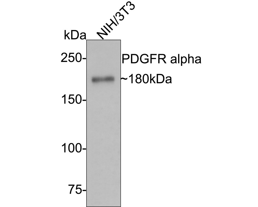
Western blot analysis of PDGFR alpha on NIH/3T3 cell lysates with Rabbit anti-PDGFR alpha antibody at 1/5000 dilution. Lysates/proteins at 10 µg/Lane. Exposure time: 2 minutes; 6% SDS-PAGE gel. Proteins were transferred to a PVDF membrane and blocked with 5% NFDM/TBST for 1 hour at room temperature. The primary antibody at 1/5000 dilution was used in 5% NFDM/TBST at room temperature for 2 hours. Goat Anti-Rabbit IgG - HRP Secondary Antibody at 1/50000 dilution was used for 1 hour at room temperature.WB
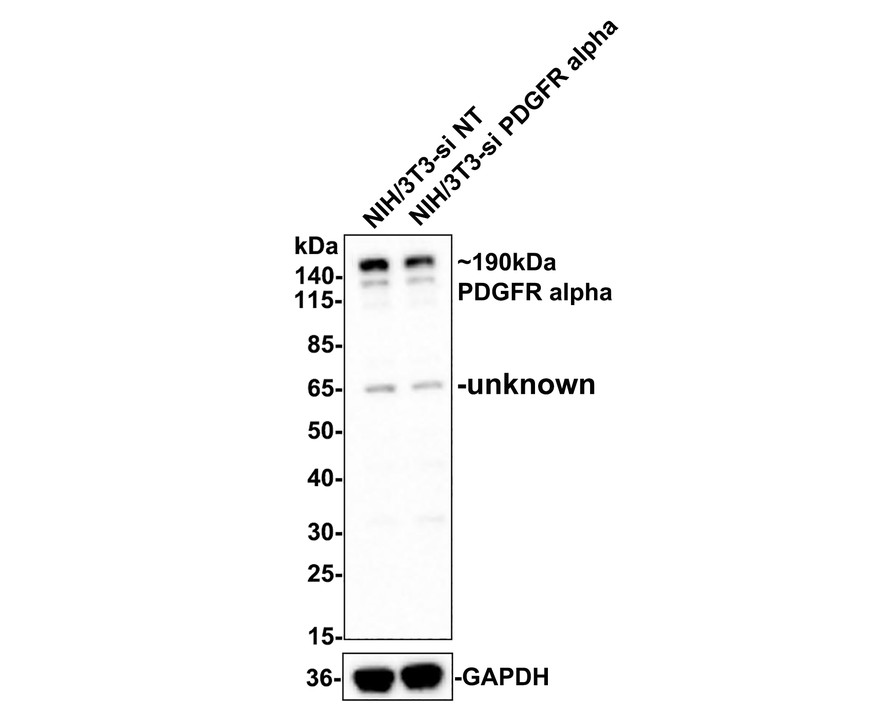
Western blot analysis of PDGFR alpha on different lysates with Rabbit anti-PDGFR alpha antibody at 1/5000 dilution. Lane 1: NIH/3T3-si NT cell lysate, Lane 2: NIH/3T3-si PDGFR alpha cell lysate, Lysates/proteins at 10 µg/Lane. Exposure time: 2 minutes; 4-20% SDS-PAGE gel. Proteins were transferred to a PVDF membrane and blocked with 5% NFDM in TBST for 1 hour at room temperature. The primary antibody (1/5000) and Loading control antibody (Rabbit anti-GAPDH,1/10000) were used in 5% BSA at room temperature for 2 hours. Goat Anti-rabbit IgG-HRP Secondary Antibody at 1/50000 dilution was used for 1 hour at room temperature.IF-F
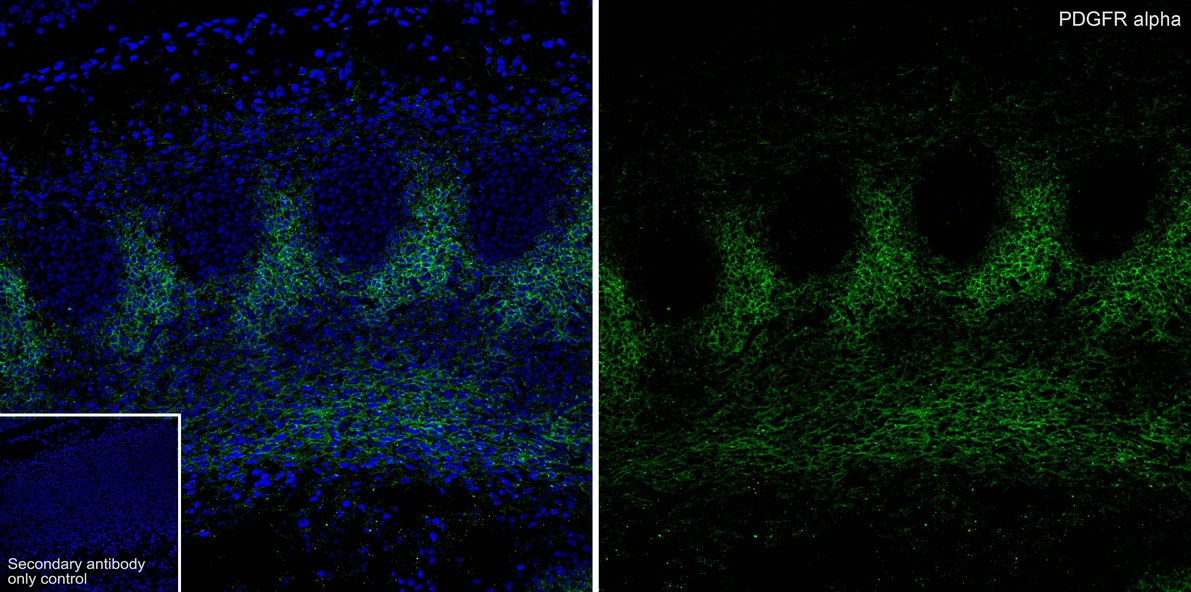
Application: IHC-Fr, Species: Mouse, Site: E14.5 embyro, Sample: Frozen section, Antibody concentration: 1:500, Antigen retrieval: Not required.| Product Name | PDGFR alpha Recombinant Rabbit Monoclonal Antibody |
|---|---|
| Antibody Type | Primary Antibodies |
| Immunogen | Synthetic peptide within C-terminal human PDGFR alpha. |
| Clonality | Monoclonal |
|---|---|
| Isotype | IgG |
| Host Species | Rabbit |
| Tested Applications | IF-FWB |
| WB:1:5000-1:10000 IF-F:1:500 |
|
| Species Reactivity | HumanMouseRat |
| Concentration | 1mg/ml |
| Purification | Affinity purified |
| Gene Symbol | PDGFRA |
|---|---|
| Gene Synonyms | CD140A PDGFR2 PDGFR-2 |
| Gene Full Name | platelet derived growth factor receptor alpha |
| Gene Summary | This gene encodes a cell surface tyrosine kinase receptor for members of the platelet-derived growth factor family. These growth factors are mitogens for cells of mesenchymal origin. The identity of the growth factor bound to a receptor monomer determines whether the functional receptor is a homodimer or a heterodimer, composed of both platelet-derived growth factor receptor alpha and beta polypeptides. Studies suggest that this gene plays a role in organ development, wound healing, and tumor progression. Mutations in this gene have been associated with idiopathic hypereosinophilic syndrome, somatic and familial gastrointestinal stromal tumors, and a variety of other cancers. [provided by RefSeq, Mar 2012] |
| Alternative Names | Alpha-type platelet-derived growth factor receptor antibody CD140 antigen-like family member A antibody CD140a antibody CD140a antigen antibody MGC74795 antibody PDGF alpha chain antibody PDGF-R-alpha antibody PDGFR 2 antibody PDGFR alpha antibody PDGFR2 antibody Alpha-type platelet-derived growth factor receptor antibody CD140 antigen-like family member A antibody CD140a antibody CD140a antigen antibody MGC74795 antibody PDGF alpha chain antibody PDGF-R-alpha antibody PDGFR 2 antibody PDGFR alpha antibody PDGFR2 antibody PDGFRA antibody PDGFRA/BCR fusion antibody PGFRA_HUMAN antibody Platelet derived growth factor receptor 2 antibody Platelet derived growth factor receptor alpha antibody Platelet derived growth factor receptor alpha polypeptide antibody Platelet derived growth factor receptor antibody Rearranged in hypereosinophilia platelet derived growth factor receptor alpha fusion protein antibody RHEPDGFRA antibody |
| Molecular Weight(MW) | 123kDa(Observed band size:180kDa) |
| Cellular Localization | Golgi apparatus, Cell membrane, cilium. |
WB
Western blot analysis of PDGFR alpha on NIH/3T3 cell lysates with Rabbit anti-PDGFR alpha antibody at 1/5000 dilution. Lysates/proteins at 10 µg/Lane. Exposure time: 2 minutes; 6% SDS-PAGE gel. Proteins were transferred to a PVDF membrane and blocked with 5% NFDM/TBST for 1 hour at room temperature. The primary antibody at 1/5000 dilution was used in 5% NFDM/TBST at room temperature for 2 hours. Goat Anti-Rabbit IgG - HRP Secondary Antibody at 1/50000 dilution was used for 1 hour at room temperature.
WB
Western blot analysis of PDGFR alpha on different lysates with Rabbit anti-PDGFR alpha antibody at 1/5000 dilution. Lane 1: NIH/3T3-si NT cell lysate, Lane 2: NIH/3T3-si PDGFR alpha cell lysate, Lysates/proteins at 10 µg/Lane. Exposure time: 2 minutes; 4-20% SDS-PAGE gel. Proteins were transferred to a PVDF membrane and blocked with 5% NFDM in TBST for 1 hour at room temperature. The primary antibody (1/5000) and Loading control antibody (Rabbit anti-GAPDH,1/10000) were used in 5% BSA at room temperature for 2 hours. Goat Anti-rabbit IgG-HRP Secondary Antibody at 1/50000 dilution was used for 1 hour at room temperature.
IF-F
Application: IHC-Fr, Species: Mouse, Site: E14.5 embyro, Sample: Frozen section, Antibody concentration: 1:500, Antigen retrieval: Not required.| Application Notes | WB:1:5000-1:10000 IF-F:1:500 |
|---|
| Form | Liquid |
|---|---|
| Storage Instructions | Shipped at 4℃. Store at +4℃ short term (1-2 weeks). It is recommended to aliquot into single-use upon delivery. Store at -20℃ long term. |
| Storage Buffer | 1*TBS (pH7.4), 0.05% BSA, 40% Glycerol. Preservative: 0.05% Sodium Azide. |


