WB
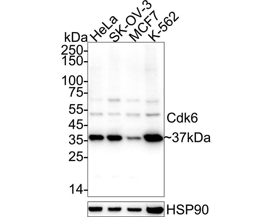
Western blot analysis of Cdk6 on different lysates with Rabbit anti-Cdk6 antibody at 1/2000 dilution. Lane 1: HeLa cell lysate, Lane 2: SK-OV-3 cell lysate, Lane 3: MCF7 cell lysate, Lane 4: K-562 cell lysate, Lysates/proteins at 20 µg/Lane. Exposure time: 59 seconds; 4-20% SDS-PAGE gel. Proteins were transferred to a PVDF membrane and blocked with 5% NFDM/TBST for 1 hour at room temperature. The primary antibody at 1/2000 dilution was used in primary antibody dilution at 4℃ overnight. Goat Anti-Rabbit IgG - HRP Secondary Antibody at 1/50000 dilution was used for 1 hour at room temperature.ICC/IF
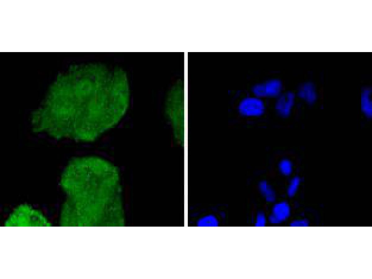
ICC staining of Cdk6 in Hela cells (green). Formalin fixed cells were permeabilized with 0.1% Triton X-100 in TBS for 10 minutes at room temperature and blocked with 1% Blocker BSA for 15 minutes at room temperature. Cells were probed with the primary antibody (1/50) for 1 hour at room temperature, washed with PBS. Alexa Fluor®488 Goat anti-Rabbit IgG was used as the secondary antibody at 1/1000 dilution. The nuclear counter stain is DAPI (blue).ICC/IF
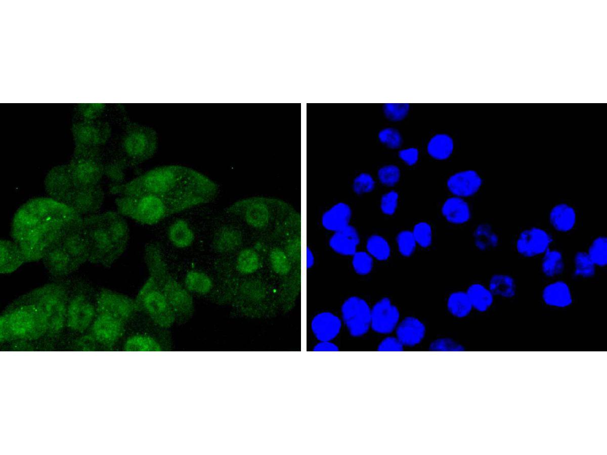
ICC staining of Cdk6 in SW480 cells (green). Formalin fixed cells were permeabilized with 0.1% Triton X-100 in TBS for 10 minutes at room temperature and blocked with 1% Blocker BSA for 15 minutes at room temperature. Cells were probed with the primary antibody (1/50) for 1 hour at room temperature, washed with PBS. Alexa Fluor®488 Goat anti-Rabbit IgG was used as the secondary antibody at 1/1,000 dilution. The nuclear counter stain is DAPI (blue).ICC/IF
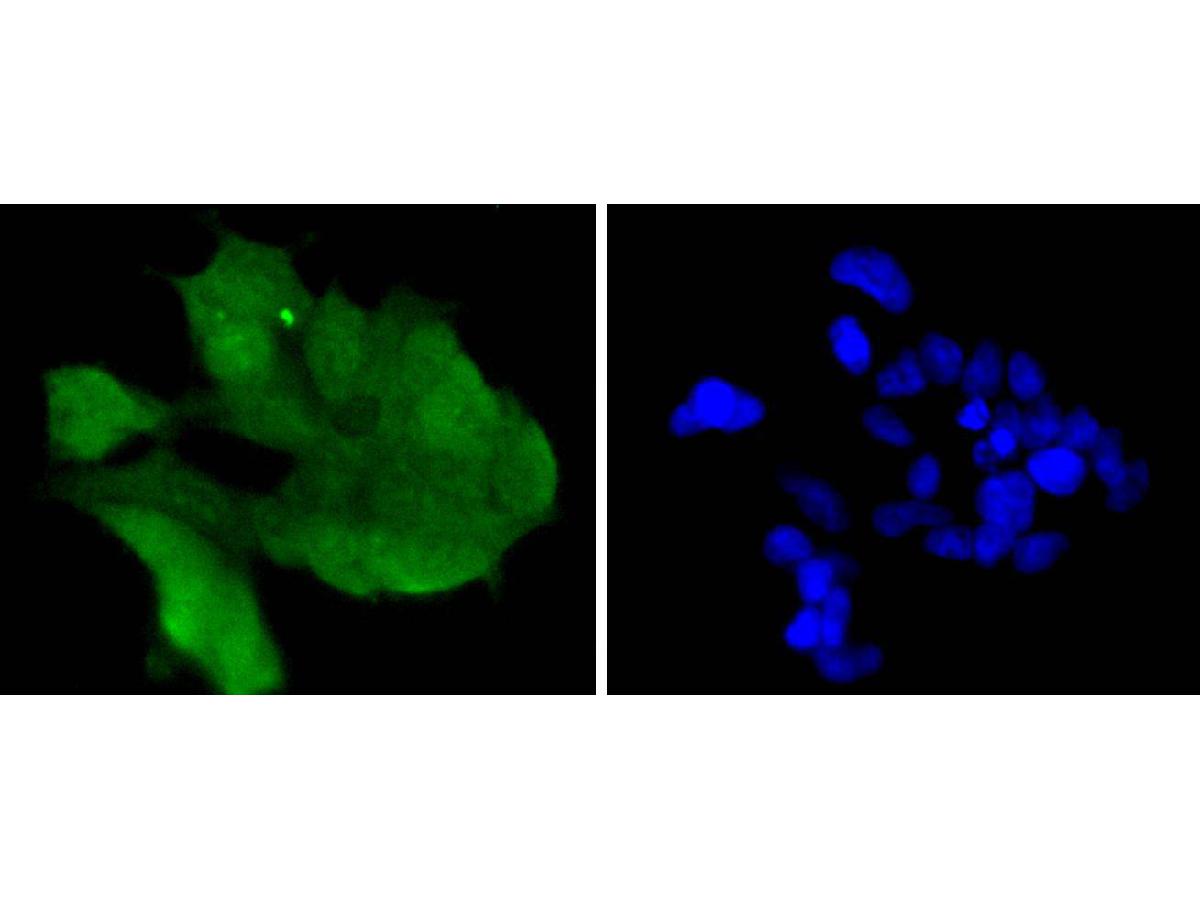
ICC staining of Cdk6 in 293 cells (green). Formalin fixed cells were permeabilized with 0.1% Triton X-100 in TBS for 10 minutes at room temperature and blocked with 1% Blocker BSA for 15 minutes at room temperature. Cells were probed with the primary antibody (1/50) for 1 hour at room temperature, washed with PBS. Alexa Fluor®488 Goat anti-Rabbit IgG was used as the secondary antibody at 1/1,000 dilution. The nuclear counter stain is DAPI (blue).IHC
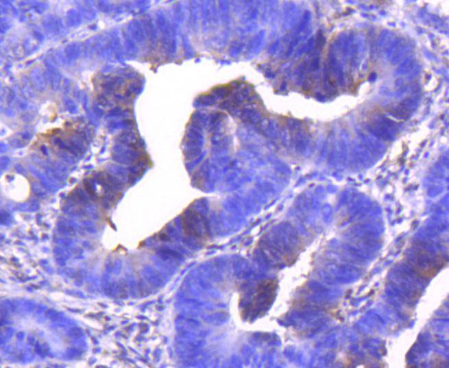
Immunohistochemical analysis of paraffin-embedded human colon carcinoma tissue using anti-Cdk6 antibody. The section was pre-treated using heat mediated antigen retrieval with Tris-EDTA buffer (pH 8.0-8.4) for 20 minutes.The tissues were blocked in 5% BSA for 30 minutes at room temperature, washed with ddH2O and PBS, and then probed with the primary antibody (1/50) for 30 minutes at room temperature. The detection was performed using an HRP conjugated compact polymer system. DAB was used as the chromogen. Tissues were counterstained with hematoxylin and mounted with DPX.FC
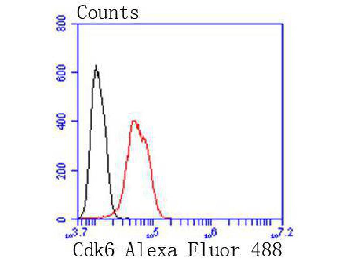
Flow cytometric analysis of Cdk6 was done on Hela cells. The cells were fixed, permeabilized and stained with the primary antibody (1/50) (red). After incubation of the primary antibody at room temperature for an hour, the cells were stained with a Alexa Fluor 488-conjugated Goat anti-Rabbit IgG Secondary antibody at 1/1000 dilution for 30 minutes.Unlabelled sample was used as a control (cells without incubation with primary antibody; black).| Product Name | Cdk6 Recombinant Rabbit Monoclonal Antibody |
|---|---|
| Antibody Type | Primary Antibodies |
| Immunogen | Synthetic peptide within Human Cdk6 aa 277-326 / 326. |
| Clonality | Monoclonal |
|---|---|
| Isotype | IgG |
| Host Species | Rabbit |
| Tested Applications | FCICC/IFIHCWB |
| WB:1:500-1:2000 ICC/IF:1:50-1:200 IHC:1:50-1:200 FC: 1:50-1:100 |
|
| Species Reactivity | Human |
| Concentration | 1mg/ml |
| Purification | Affinity purified |
| Gene Symbol | CDK6 |
|---|---|
| Gene Synonyms | MCPH12 PLSTIRE |
| Gene Full Name | cyclin dependent kinase 6 |
| Gene Summary | The protein encoded by this gene is a member of the CMGC family of serine/threonine protein kinases. This kinase is a catalytic subunit of the protein kinase complex that is important for cell cycle G1 phase progression and G1/S transition. The activity of this kinase first appears in mid-G1 phase, which is controlled by the regulatory subunits including D-type cyclins and members of INK4 family of CDK inhibitors. This kinase, as well as CDK4, has been shown to phosphorylate, and thus regulate the activity of, tumor suppressor protein Rb. Altered expression of this gene has been observed in multiple human cancers. A mutation in this gene resulting in reduced cell proliferation, and impaired cell motility and polarity, and has been identified in patients with primary microcephaly. [provided by RefSeq, Aug 2017] |
| Alternative Names | CDK 6 antibody CDK6 antibody CDK6_HUMAN antibody Cell division protein kinase 6 antibody Crk 2 antibody Crk2 antibody Cyclin dependent kinase 6 antibody Cyclin-dependent kinase 6 antibody MGC59692 antibody p40 antibody CDK 6 antibody CDK6 antibody CDK6_HUMAN antibody Cell division protein kinase 6 antibody Crk 2 antibody Crk2 antibody Cyclin dependent kinase 6 antibody Cyclin-dependent kinase 6 antibody MGC59692 antibody p40 antibody PLSTIRE antibody Serine/threonine protein kinase PLSTIRE antibody Serine/threonine-protein kinase PLSTIRE antibody STQTL11 antibody |
| Molecular Weight(MW) | 37kDa(Observed band size:37kDa) |
| Cellular Localization | Cytoplasm, Nucleus, Cell projection. |
WB
Western blot analysis of Cdk6 on different lysates with Rabbit anti-Cdk6 antibody at 1/2000 dilution. Lane 1: HeLa cell lysate, Lane 2: SK-OV-3 cell lysate, Lane 3: MCF7 cell lysate, Lane 4: K-562 cell lysate, Lysates/proteins at 20 µg/Lane. Exposure time: 59 seconds; 4-20% SDS-PAGE gel. Proteins were transferred to a PVDF membrane and blocked with 5% NFDM/TBST for 1 hour at room temperature. The primary antibody at 1/2000 dilution was used in primary antibody dilution at 4℃ overnight. Goat Anti-Rabbit IgG - HRP Secondary Antibody at 1/50000 dilution was used for 1 hour at room temperature.
ICC/IF
ICC staining of Cdk6 in Hela cells (green). Formalin fixed cells were permeabilized with 0.1% Triton X-100 in TBS for 10 minutes at room temperature and blocked with 1% Blocker BSA for 15 minutes at room temperature. Cells were probed with the primary antibody (1/50) for 1 hour at room temperature, washed with PBS. Alexa Fluor®488 Goat anti-Rabbit IgG was used as the secondary antibody at 1/1000 dilution. The nuclear counter stain is DAPI (blue).
ICC/IF
ICC staining of Cdk6 in SW480 cells (green). Formalin fixed cells were permeabilized with 0.1% Triton X-100 in TBS for 10 minutes at room temperature and blocked with 1% Blocker BSA for 15 minutes at room temperature. Cells were probed with the primary antibody (1/50) for 1 hour at room temperature, washed with PBS. Alexa Fluor®488 Goat anti-Rabbit IgG was used as the secondary antibody at 1/1,000 dilution. The nuclear counter stain is DAPI (blue).
ICC/IF
ICC staining of Cdk6 in 293 cells (green). Formalin fixed cells were permeabilized with 0.1% Triton X-100 in TBS for 10 minutes at room temperature and blocked with 1% Blocker BSA for 15 minutes at room temperature. Cells were probed with the primary antibody (1/50) for 1 hour at room temperature, washed with PBS. Alexa Fluor®488 Goat anti-Rabbit IgG was used as the secondary antibody at 1/1,000 dilution. The nuclear counter stain is DAPI (blue).
IHC
Immunohistochemical analysis of paraffin-embedded human colon carcinoma tissue using anti-Cdk6 antibody. The section was pre-treated using heat mediated antigen retrieval with Tris-EDTA buffer (pH 8.0-8.4) for 20 minutes.The tissues were blocked in 5% BSA for 30 minutes at room temperature, washed with ddH2O and PBS, and then probed with the primary antibody (1/50) for 30 minutes at room temperature. The detection was performed using an HRP conjugated compact polymer system. DAB was used as the chromogen. Tissues were counterstained with hematoxylin and mounted with DPX.
FC
Flow cytometric analysis of Cdk6 was done on Hela cells. The cells were fixed, permeabilized and stained with the primary antibody (1/50) (red). After incubation of the primary antibody at room temperature for an hour, the cells were stained with a Alexa Fluor 488-conjugated Goat anti-Rabbit IgG Secondary antibody at 1/1000 dilution for 30 minutes.Unlabelled sample was used as a control (cells without incubation with primary antibody; black).| Application Notes | WB:1:500-1:2000 ICC/IF:1:50-1:200 IHC:1:50-1:200 FC: 1:50-1:100 |
|---|
| Form | Liquid |
|---|---|
| Storage Instructions | Shipped at 4℃. Store at +4℃ short term (1-2 weeks). It is recommended to aliquot into single-use upon delivery. Store at -20℃ long term. |
| Storage Buffer | 1*TBS (pH7.4), 0.05% BSA, 40% Glycerol. Preservative: 0.05% Sodium Azide. |


