WB
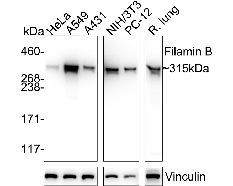
Western blot analysis of Filamin B on different lysates with Rabbit anti-Filamin B antibody at 1/5000 dilution. Lane 1: HeLa cell lysate (20 µg/Lane), Lane 2: A549 cell lysate (20 µg/Lane), Lane 3: A431 cell lysate (20 µg/Lane), Lane 4: NIH/3T3 cell lysate (20 µg/Lane), Lane 5: PC-12 cell lysate (20 µg/Lane), Lane 6: Rat lung tissue lysate (40 µg/Lane), Exposure time: Lane 1-3: 4 seconds; Lane 4-6: 42 seconds; 3-8% SDS-PAGE gel. Proteins were transferred to a PVDF membrane and blocked with 5% NFDM/TBST for 1 hour at room temperature. The primary antibody at 1/5000 dilution was used in 5% NFDM/TBST at 4℃ overnight. Goat Anti-Rabbit IgG - HRP Secondary Antibody at 1/50000 dilution was used for 1 hour at room temperature.IHC
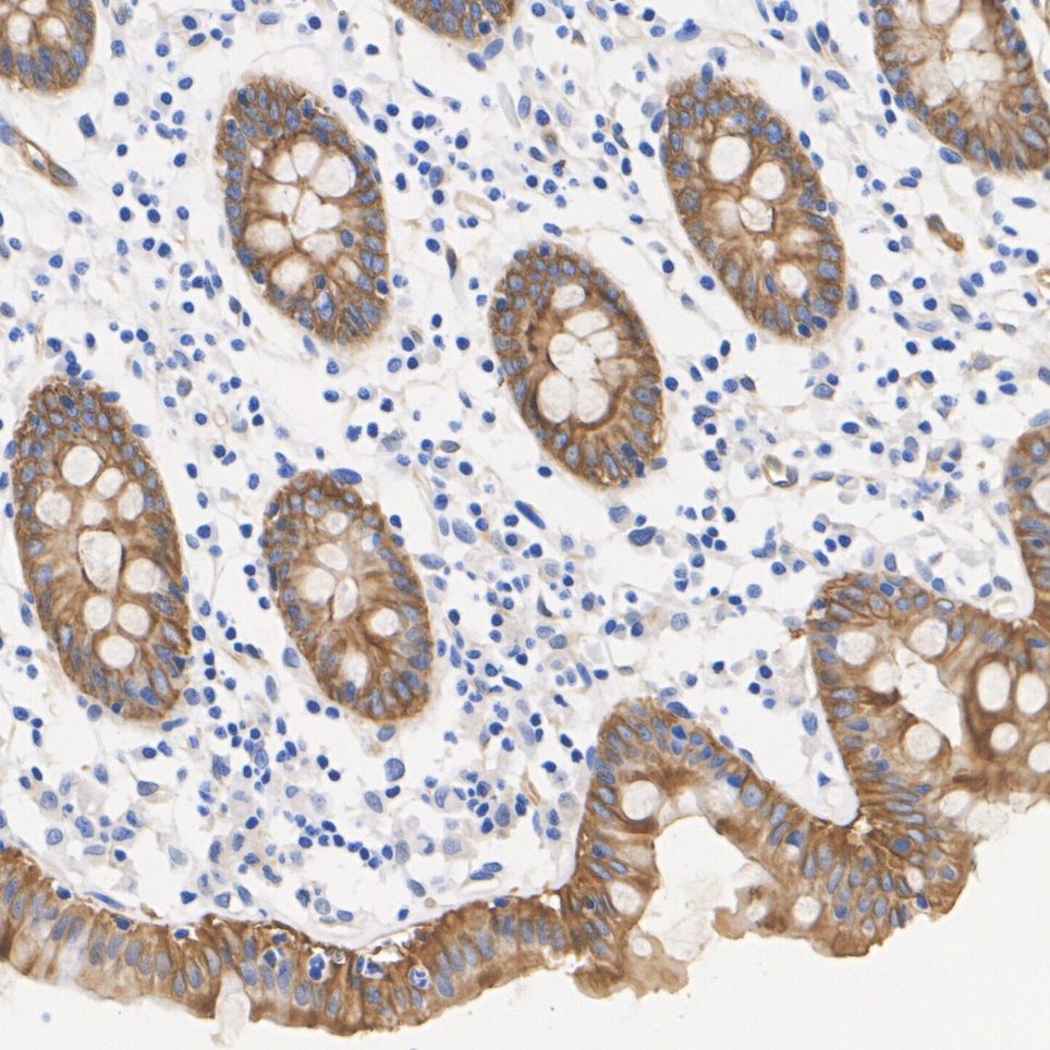
Immunohistochemical analysis of paraffin-embedded human colon tissue with Rabbit anti-Filamin B antibody at 1/5000 dilution. The section was pre-treated using heat mediated antigen retrieval with Tris-EDTA buffer (pH 9.0) for 20 minutes. The tissues were blocked in 1% BSA for 20 minutes at room temperature, washed with ddH2O and PBS, and then probed with the primary antibody at 1/5000 dilution for 1 hour at room temperature. The detection was performed using an HRP conjugated compact polymer system. DAB was used as the chromogen. Tissues were counterstained with hematoxylin and mounted with DPX.IHC
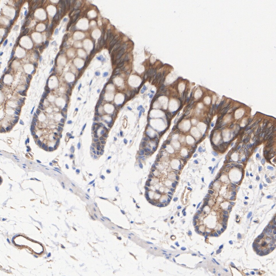
Immunohistochemical analysis of paraffin-embedded rat colon tissue with Rabbit anti-Filamin B antibody at 1/5000 dilution. The section was pre-treated using heat mediated antigen retrieval with Tris-EDTA buffer (pH 9.0) for 20 minutes. The tissues were blocked in 1% BSA for 20 minutes at room temperature, washed with ddH2O and PBS, and then probed with the primary antibody at 1/5000 dilution for 1 hour at room temperature. The detection was performed using an HRP conjugated compact polymer system. DAB was used as the chromogen. Tissues were counterstained with hematoxylin and mounted with DPX.| Product Name | Filamin B Recombinant Rabbit Monoclonal Antibody |
|---|---|
| Antibody Type | Primary Antibodies |
| Immunogen | Recombinant protein within human Filamin B aa |
| Clonality | Monoclonal |
|---|---|
| Isotype | IgG |
| Host Species | Rabbit |
| Tested Applications | IHCWB |
| WB:1:5000 IHC:1:5000 |
|
| Species Reactivity | HumanMouseRat |
| Concentration | 1mg/ml |
| Purification | Affinity purified |
| Gene Symbol | FLNB |
|---|---|
| Gene Synonyms | FH1 TAP LRS1 TABP FLN-B FLN1L ABP-278 ABP-280 |
| Gene Full Name | filamin B |
| Gene Summary | This gene encodes a member of the filamin family. The encoded protein interacts with glycoprotein Ib alpha as part of the process to repair vascular injuries. The platelet glycoprotein Ib complex includes glycoprotein Ib alpha, and it binds the actin cytoskeleton. Mutations in this gene have been found in several conditions: atelosteogenesis type 1 and type 3; boomerang dysplasia; autosomal dominant Larsen syndrome; and spondylocarpotarsal synostosis syndrome. Multiple alternatively spliced transcript variants that encode different protein isoforms have been described for this gene. [provided by RefSeq, Nov 2009] |
| Alternative Names | ABP 278 antibody ABP 280 homolog antibody ABP-278 antibody ABP-280 homolog antibody ABP278 antibody Actin binding like protein antibody Actin binding protein 278 antibody Actin-binding-like protein antibody AOI antibody Beta filamin antibody ABP 278 antibody ABP 280 homolog antibody ABP-278 antibody ABP-280 homolog antibody ABP278 antibody Actin binding like protein antibody Actin binding protein 278 antibody Actin-binding-like protein antibody AOI antibody Beta filamin antibody Beta-filamin antibody DKFZp686A1668 antibody DKFZp686O033 antibody Fh1 antibody Filamin 1 actin binding protein 280 like antibody Filamin 3 antibody Filamin B antibody Filamin B beta actin binding protein 278 antibody Filamin homolog 1 antibody Filamin-3 antibody Filamin-B antibody FLN B antibody FLN-B antibody FLN1L antibody FLN3 antibody FLNB antibody FLNB_HUMAN antibody LRS1 antibody SCT antibody TABP antibody TAP antibody Thyroid autoantigen antibody Truncated ABP antibody Truncated actin binding protein antibody Truncated actin-binding protein antibody |
| Molecular Weight(MW) | 278kDa(Observed band size:315kDa) |
| Cellular Localization | Cytoplasm, cell cortex, cytoskeleton, stress, myofibril, sarcomere, Z line. |
WB
Western blot analysis of Filamin B on different lysates with Rabbit anti-Filamin B antibody at 1/5000 dilution. Lane 1: HeLa cell lysate (20 µg/Lane), Lane 2: A549 cell lysate (20 µg/Lane), Lane 3: A431 cell lysate (20 µg/Lane), Lane 4: NIH/3T3 cell lysate (20 µg/Lane), Lane 5: PC-12 cell lysate (20 µg/Lane), Lane 6: Rat lung tissue lysate (40 µg/Lane), Exposure time: Lane 1-3: 4 seconds; Lane 4-6: 42 seconds; 3-8% SDS-PAGE gel. Proteins were transferred to a PVDF membrane and blocked with 5% NFDM/TBST for 1 hour at room temperature. The primary antibody at 1/5000 dilution was used in 5% NFDM/TBST at 4℃ overnight. Goat Anti-Rabbit IgG - HRP Secondary Antibody at 1/50000 dilution was used for 1 hour at room temperature.
IHC
Immunohistochemical analysis of paraffin-embedded human colon tissue with Rabbit anti-Filamin B antibody at 1/5000 dilution. The section was pre-treated using heat mediated antigen retrieval with Tris-EDTA buffer (pH 9.0) for 20 minutes. The tissues were blocked in 1% BSA for 20 minutes at room temperature, washed with ddH2O and PBS, and then probed with the primary antibody at 1/5000 dilution for 1 hour at room temperature. The detection was performed using an HRP conjugated compact polymer system. DAB was used as the chromogen. Tissues were counterstained with hematoxylin and mounted with DPX.
IHC
Immunohistochemical analysis of paraffin-embedded rat colon tissue with Rabbit anti-Filamin B antibody at 1/5000 dilution. The section was pre-treated using heat mediated antigen retrieval with Tris-EDTA buffer (pH 9.0) for 20 minutes. The tissues were blocked in 1% BSA for 20 minutes at room temperature, washed with ddH2O and PBS, and then probed with the primary antibody at 1/5000 dilution for 1 hour at room temperature. The detection was performed using an HRP conjugated compact polymer system. DAB was used as the chromogen. Tissues were counterstained with hematoxylin and mounted with DPX.| Application Notes | WB:1:5000 IHC:1:5000 |
|---|
| Form | Liquid |
|---|---|
| Storage Instructions | Shipped at 4℃. Store at +4℃ short term (1-2 weeks). It is recommended to aliquot into single-use upon delivery. Store at -20℃ long term. |
| Storage Buffer | PBS (pH7.4), 0.1% BSA, 40% Glycerol. Preservative: 0.05% Sodium Azide. |


