WB
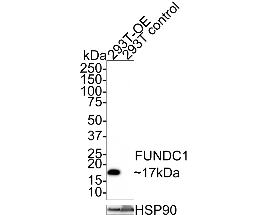
Western blot analysis of FUNDC1 on different lysates with Rabbit anti-FUNDC1 antibody at 1/1000 dilution. Lane 1: 293T over-expressed FUNDC1 cell lysate, Lane 2: 293T control cell lysate, Lysates/proteins at 20 µg/Lane. Exposure time: 2 minutes; 4-20% SDS-PAGE gel. Proteins were transferred to a PVDF membrane and blocked with 5% NFDM/TBST for 1 hour at room temperature. The primary antibody at 1/1000 dilution was used in 5% NFDM/TBST at room temperature for 2 hours. Goat Anti-Rabbit IgG - HRP Secondary Antibody at 1:100000 dilution was used for 1 hour at room temperature.WB
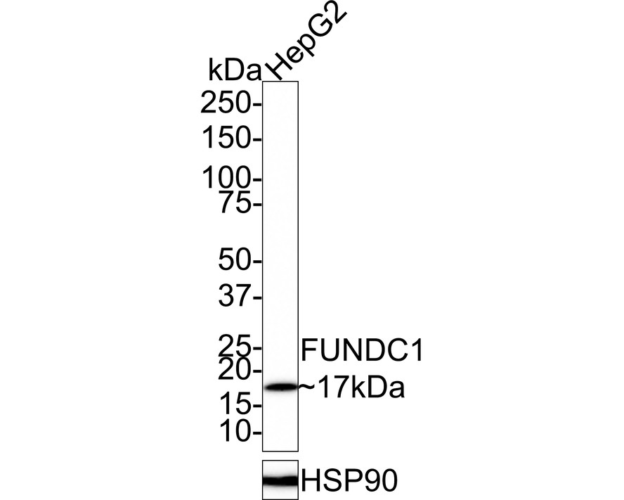
Western blot analysis of FUNDC1 on HepG2 cell lysates with Rabbit anti-FUNDC1 antibody at 1/1000 dilution. Lysates/proteins at 40 µg/Lane. Exposure time: 2 minutes; 4-20% SDS-PAGE gel. Proteins were transferred to a PVDF membrane and blocked with 5% NFDM/TBST for 1 hour at room temperature. The primary antibody at 1/1000 dilution was used in 5% NFDM/TBST at 4℃ overnight. Goat Anti-Rabbit IgG - HRP Secondary Antibody at 1:100000 dilution was used for 1 hour at room temperature.IHC
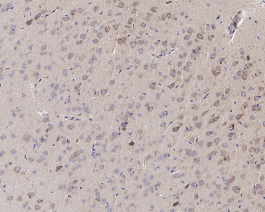
Immunohistochemical analysis of paraffin-embedded rat brain tissue with Rabbit anti-FUNDC1 antibody at 1/50 dilution. The section was pre-treated using heat mediated antigen retrieval with Tris-EDTA buffer (pH 9.0) for 20 minutes. The tissues were blocked in 1% BSA for 20 minutes at room temperature, washed with ddH2O and PBS, and then probed with the primary antibody at 1/50 dilution for 1 hour at room temperature. The detection was performed using an HRP conjugated compact polymer system. DAB was used as the chromogen. Tissues were counterstained with hematoxylin and mounted with DPX.| Product Name | FUNDC1 Recombinant Rabbit Monoclonal Antibody |
|---|---|
| Antibody Type | Primary Antibodies |
| Immunogen | Recombinant protein within N terminal human FUNDC1. |
| Clonality | Monoclonal |
|---|---|
| Isotype | IgG |
| Host Species | Rabbit |
| Tested Applications | IHCWB |
| WB:1:1000 IHC:1:50 |
|
| Species Reactivity | HumanRat |
| Concentration | 1mg/ml |
| Purification | Affinity purified |
| Gene Symbol | FUNDC1 |
|---|---|
| Gene Full Name | FUN14 domain containing 1 |
| Gene Summary | This gene encodes a protein with a FUN14 superfamily domain. The function of the encoded protein is not known. [provided by RefSeq, Sep 2011] |
| Alternative Names | FUN14 domain containing 1 antibody FUN14 domain containing protein 1 antibody FUN14 domain-containing protein 1 antibody FUND1_HUMAN antibody fundc1 antibody |
| Molecular Weight(MW) | 17kDa(Observed band size:17kDa) |
| Cellular Localization | Mitochondrion outer membrane. |
WB
Western blot analysis of FUNDC1 on different lysates with Rabbit anti-FUNDC1 antibody at 1/1000 dilution. Lane 1: 293T over-expressed FUNDC1 cell lysate, Lane 2: 293T control cell lysate, Lysates/proteins at 20 µg/Lane. Exposure time: 2 minutes; 4-20% SDS-PAGE gel. Proteins were transferred to a PVDF membrane and blocked with 5% NFDM/TBST for 1 hour at room temperature. The primary antibody at 1/1000 dilution was used in 5% NFDM/TBST at room temperature for 2 hours. Goat Anti-Rabbit IgG - HRP Secondary Antibody at 1:100000 dilution was used for 1 hour at room temperature.
WB
Western blot analysis of FUNDC1 on HepG2 cell lysates with Rabbit anti-FUNDC1 antibody at 1/1000 dilution. Lysates/proteins at 40 µg/Lane. Exposure time: 2 minutes; 4-20% SDS-PAGE gel. Proteins were transferred to a PVDF membrane and blocked with 5% NFDM/TBST for 1 hour at room temperature. The primary antibody at 1/1000 dilution was used in 5% NFDM/TBST at 4℃ overnight. Goat Anti-Rabbit IgG - HRP Secondary Antibody at 1:100000 dilution was used for 1 hour at room temperature.
IHC
Immunohistochemical analysis of paraffin-embedded rat brain tissue with Rabbit anti-FUNDC1 antibody at 1/50 dilution. The section was pre-treated using heat mediated antigen retrieval with Tris-EDTA buffer (pH 9.0) for 20 minutes. The tissues were blocked in 1% BSA for 20 minutes at room temperature, washed with ddH2O and PBS, and then probed with the primary antibody at 1/50 dilution for 1 hour at room temperature. The detection was performed using an HRP conjugated compact polymer system. DAB was used as the chromogen. Tissues were counterstained with hematoxylin and mounted with DPX.| Application Notes | WB:1:1000 IHC:1:50 |
|---|
| Form | Liquid |
|---|---|
| Storage Instructions | Shipped at 4℃. Store at +4℃ short term (1-2 weeks). It is recommended to aliquot into single-use upon delivery. Store at -20℃ long term. |
| Storage Buffer | PBS (pH7.4), 0.1% BSA, 40% Glycerol. Preservative: 0.05% Sodium Azide. |


