WB
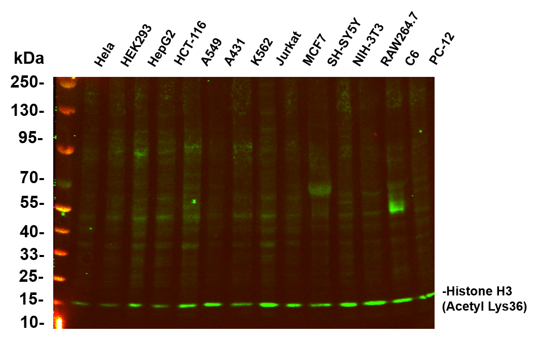
Various whole cell lysates were separated by 4-20% SDS-PAGE, and the primary antibody was used at 4℃, over night with a 1:5000 dilution . The Dylight 800-conjugated Goat anti-Rabbit antibody was used to detect the antibody. Lane1: Hela - Human cervical cancer Lane2: HEK293 - Human normal embryonic kidney Lane3: HepG2 - Human hepatocellular carcinoma Lane4: HCT-116 - Human colon cancer Lane5: A549 - Human lung adenocarcinoma Lane6: A431 - Human skin squamous cell carcinoma Lane7: K562 - Human chronic myeloid leukemia Lane8: Jurkat - Human acute T cell leukemia cells Lane9: MCF7 - Human breast cancer Lane10: SH-SY5Y - Human neuroblastoma cells Lane11: NIH-3T3 - NIH mouse fibroblasts Lane12: RAW264.7 - Mouse mononuclear macrophage leukemia cells Lane13: C6 - Rat glioma cells Lane14: PC-12 - Pheochromocytoma in rats.WB
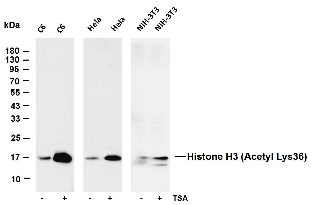
Various whole cell lysates were separated by 4-20% SDS-PAGE, and the membrane was blotted with anti-MEK1/2 (Phospho Ser217/221) antibody. The HRP-conjugated Goat anti-Rabbit IgG(H + L) antibody was used to detect the antibody. Lane 1: C6 Lane 2: C6 was treated with Trichostatin A(500 ng/mL) Lane 3: Hela Lane 4: Hela was treated with Trichostatin A overnight Lane 5: NIH-3T3 Lane 6: NIH-3T3 was treated with Trichostatin A overnight.ICC/IF
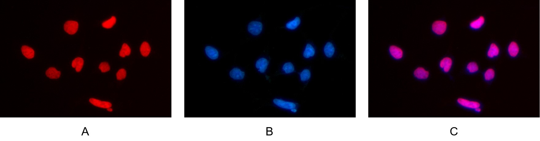
Immunofluorescence analysis of HEK293. Picture A: Histone H3 (Acetyl Lys36) antibody (red). Picture B: DAPI (blue). Picture C: Merge of A+B.IHC
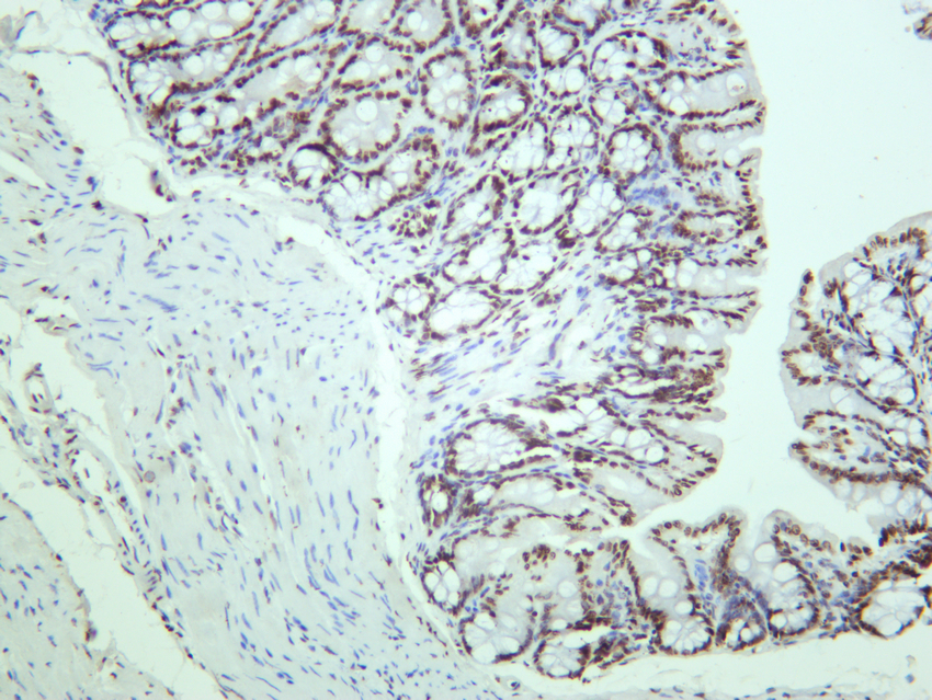
Mouse colon was stained with anti-Histone H3 (Acetyl Lys36) rabbit antibody.IHC
Rat colon was stained with anti-Histone H3 (Acetyl Lys36) rabbit antibody.IHC
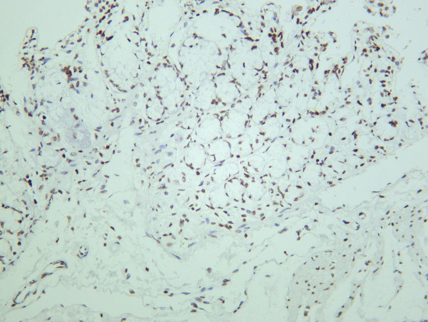
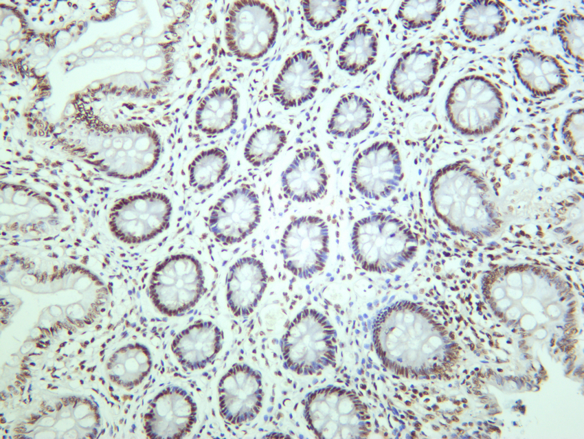
Human colon was stained with anti-Histone H3 (Acetyl Lys36) rabbit antibody.| Product Name | Histone H3 (Acetyl Lys36) Rabbit mAb |
|---|---|
| Antibody Type | Primary Antibodies |
| Clonality | monoclonal |
|---|---|
| Isotype | IgG,K |
| Host Species | Rabbit |
| Tested Applications | ICC/IFIHCWB |
| WB:1:2000-1:10000 ICC/IF:1:200-1:1000 IHC:1:200-1:1000 |
|
| Species Reactivity | HumanMouseRat |
| Concentration | 1mg/ml |
| Purification | Protein A |
| Gene Symbol | H3C1 |
|---|---|
| Gene Synonyms | H3/A H3FA HIST1H3A |
| Gene Full Name | H3 clustered histone 1 |
| Gene Summary | Histones are basic nuclear proteins that are responsible for the nucleosome structure of the chromosomal fiber in eukaryotes. This structure consists of approximately 146 bp of DNA wrapped around a nucleosome, an octamer composed of pairs of each of the four core histones (H2A, H2B, H3, and H4). The chromatin fiber is further compacted through the interaction of a linker histone, H1, with the DNA between the nucleosomes to form higher order chromatin structures. This gene is intronless and encodes a replication-dependent histone that is a member of the histone H3 family. Transcripts from this gene lack polyA tails; instead, they contain a palindromic termination element. This gene is found in the large histone gene cluster on chromosome 6p22-p21.3. [provided by RefSeq, Aug 2015] |
| Alternative Names | HIST1H3A H3FA HIST1H3B H3FL HIST1H3C H3FC HIST1H3D H3FB HIST1H3E H3FD HIST1H3F H3FI HIST1H3G H3FH HIST1H3H H3FK HIST1H3I H3FF HIST1H3J H3FJ Histone H3.1 Histone H3/a Histone H3/b Histone H3/c Histone H3/d Histone H3/f Histone H3/h Histone H3/i Histone H3/j Histone H3/k Histone H3/l HIST2H3A HIST2H3C H3F2 H3FM HIST2H3D Histone H3.2 Histone H3/m Histone H3/o H3F3A H3.3A H3F3 PP781 H3F3B H3.3B Histone H3.3 H3k36AC |
| Molecular Weight(MW) | 15kDa(Observed MW 15kDa) |
| Cellular Localization | Nucleus. Chromosome. |
WB
Various whole cell lysates were separated by 4-20% SDS-PAGE, and the primary antibody was used at 4℃, over night with a 1:5000 dilution . The Dylight 800-conjugated Goat anti-Rabbit antibody was used to detect the antibody. Lane1: Hela - Human cervical cancer Lane2: HEK293 - Human normal embryonic kidney Lane3: HepG2 - Human hepatocellular carcinoma Lane4: HCT-116 - Human colon cancer Lane5: A549 - Human lung adenocarcinoma Lane6: A431 - Human skin squamous cell carcinoma Lane7: K562 - Human chronic myeloid leukemia Lane8: Jurkat - Human acute T cell leukemia cells Lane9: MCF7 - Human breast cancer Lane10: SH-SY5Y - Human neuroblastoma cells Lane11: NIH-3T3 - NIH mouse fibroblasts Lane12: RAW264.7 - Mouse mononuclear macrophage leukemia cells Lane13: C6 - Rat glioma cells Lane14: PC-12 - Pheochromocytoma in rats.
WB
Various whole cell lysates were separated by 4-20% SDS-PAGE, and the membrane was blotted with anti-MEK1/2 (Phospho Ser217/221) antibody. The HRP-conjugated Goat anti-Rabbit IgG(H + L) antibody was used to detect the antibody. Lane 1: C6 Lane 2: C6 was treated with Trichostatin A(500 ng/mL) Lane 3: Hela Lane 4: Hela was treated with Trichostatin A overnight Lane 5: NIH-3T3 Lane 6: NIH-3T3 was treated with Trichostatin A overnight.
ICC/IF
Immunofluorescence analysis of HEK293. Picture A: Histone H3 (Acetyl Lys36) antibody (red). Picture B: DAPI (blue). Picture C: Merge of A+B.
IHC
Mouse colon was stained with anti-Histone H3 (Acetyl Lys36) rabbit antibody.
IHC
Rat colon was stained with anti-Histone H3 (Acetyl Lys36) rabbit antibody.
IHC
Human colon was stained with anti-Histone H3 (Acetyl Lys36) rabbit antibody.| Application Notes | WB:1:2000-1:10000 ICC/IF:1:200-1:1000 IHC:1:200-1:1000 |
|---|
| Form | Liquid |
|---|---|
| Storage Instructions | -15°C to -25°C/1 year(Do not lower than -25°C) |
| Storage Buffer | PBS, 50% glycerol, 0.05% Proclin 300, 0.05%BSA |


