WB
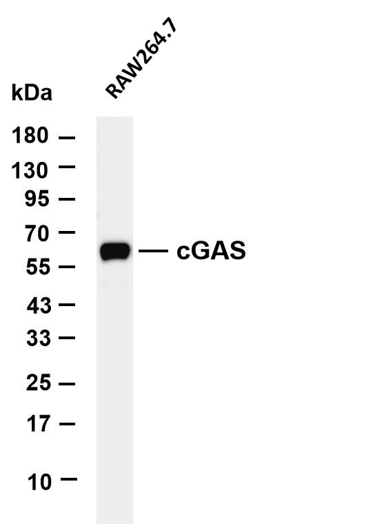
Various whole cell lysates were separated by 4-20% SDS-PAGE, and the membrane was blotted with anti-cGAS antibody. The HRP-conjugated Goat anti-Rabbit IgG(H + L) antibody was used to detect the antibody. Lane 1: RAW264.7.IHC
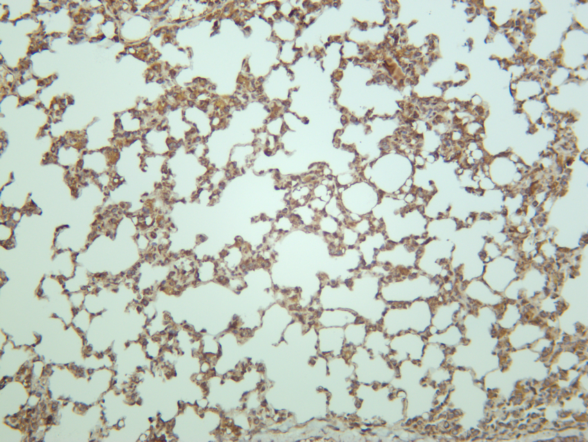
Mouse lung was stained with anti-cGAS rabbit antibody.ICC/IF
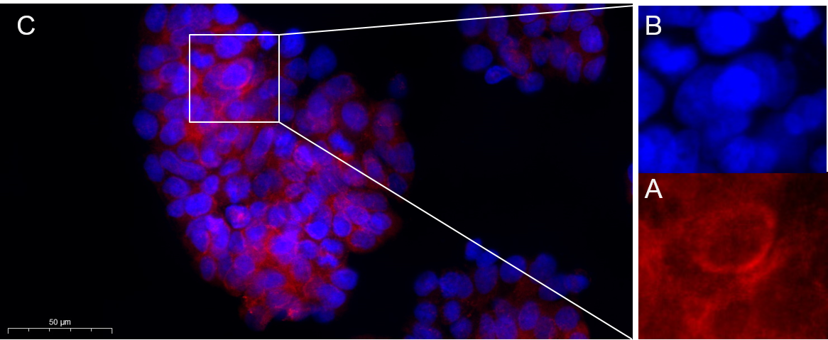
Immunofluorescence analysis of HT-29 . Picture A: cGAS Rabbit mAb (red). Picture B: DAPI (blue). Picture C: Merge of A+B.| Product Name | cGAS Rabbit mAb |
|---|---|
| Antibody Type | Primary Antibodies |
| Clonality | monoclonal |
|---|---|
| Isotype | IgG,K |
| Host Species | Rabbit |
| Tested Applications | ICC/IFIHCWB |
| WB:1:2000-1:10000 IHC:1:200-1:1000 ICC/IF:1:200-1:1000 |
|
| Species Reactivity | Mouse |
| Concentration | 1mg/ml |
| Purification | Protein A |
| Gene Symbol | CGAS |
|---|---|
| Gene Synonyms | D4 MB21D1 h-cGAS C6orf150 |
| Gene Full Name | cyclic GMP-AMP synthase |
| Gene Summary | Enables several functions, including 2',3'-cyclic GMP-AMP synthase activity; molecular condensate scaffold activity; and phosphatidylinositol-4,5-bisphosphate binding activity. Involved in several processes, including intracellular signal transduction; paracrine signaling; and regulation of defense response. Located in nuclear body; plasma membrane; and site of double-strand break. Is active in cytosol and nucleus. [provided by Alliance of Genome Resources, Jul 2025] |
| Molecular Weight(MW) | 58kDa(Observed MW 58kDa) |
| Cellular Localization | Cytoplasm, Nucleus |
WB
Various whole cell lysates were separated by 4-20% SDS-PAGE, and the membrane was blotted with anti-cGAS antibody. The HRP-conjugated Goat anti-Rabbit IgG(H + L) antibody was used to detect the antibody. Lane 1: RAW264.7.
IHC
Mouse lung was stained with anti-cGAS rabbit antibody.
ICC/IF
Immunofluorescence analysis of HT-29 . Picture A: cGAS Rabbit mAb (red). Picture B: DAPI (blue). Picture C: Merge of A+B.| Application Notes | WB:1:2000-1:10000 IHC:1:200-1:1000 ICC/IF:1:200-1:1000 |
|---|
| Form | Liquid |
|---|---|
| Storage Instructions | -15°C to -25°C/1 year(Do not lower than -25°C) |
| Storage Buffer | PBS, 50% glycerol, 0.05% Proclin 300, 0.05%BSA |


