WB
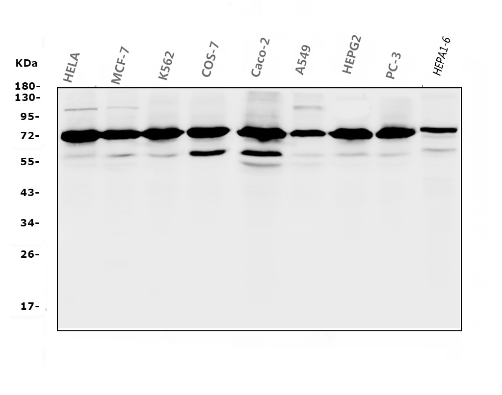
Western blot analysis of RPA1 using anti-RPA1 antibody. The sample well of each lane was loaded with 30 ug of sample under reducing conditions. Lane 1: HELA whole cell lysates, Lane 2: MCF-7 whole cell lysates, Lane 3: K562 whole cell lysates, Lane 4: COS-7 whole cell lysates, Lane 5: Caco-2 whole cell lysates, Lane 6: A549 whole cell lysates, Lane 7: HEPG2 whole cell lysates, Lane 8: PC-3 whole cell lysates, Lane 9: HEPA1-6 whole cell lysates. After electrophoresis, proteins were transferred to a membrane. Then the membrane was incubated with mouse anti-RPA1 antigen affinity purified monoclonal antibody at a dilution of 1:1000 and probed with a goat anti-mouse IgG-HRP secondary antibody. The signal is developed using ECL Plus Western Blotting Substrate.IHC
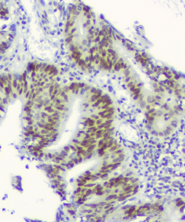
IHC analysis of RPA1 using anti-RPA1 antibody. RPA1 was detected in a paraffin-embedded section of human intestinal cancer tissue. Biotinylated goat anti-mouse IgG was used as secondary antibody. The tissue section was incubated with mouse anti-RPA1 Antibody at a dilution of 1:200 and developed using Strepavidin-Biotin-Complex (SABC) with DAB as the chromogen.ICC/IF
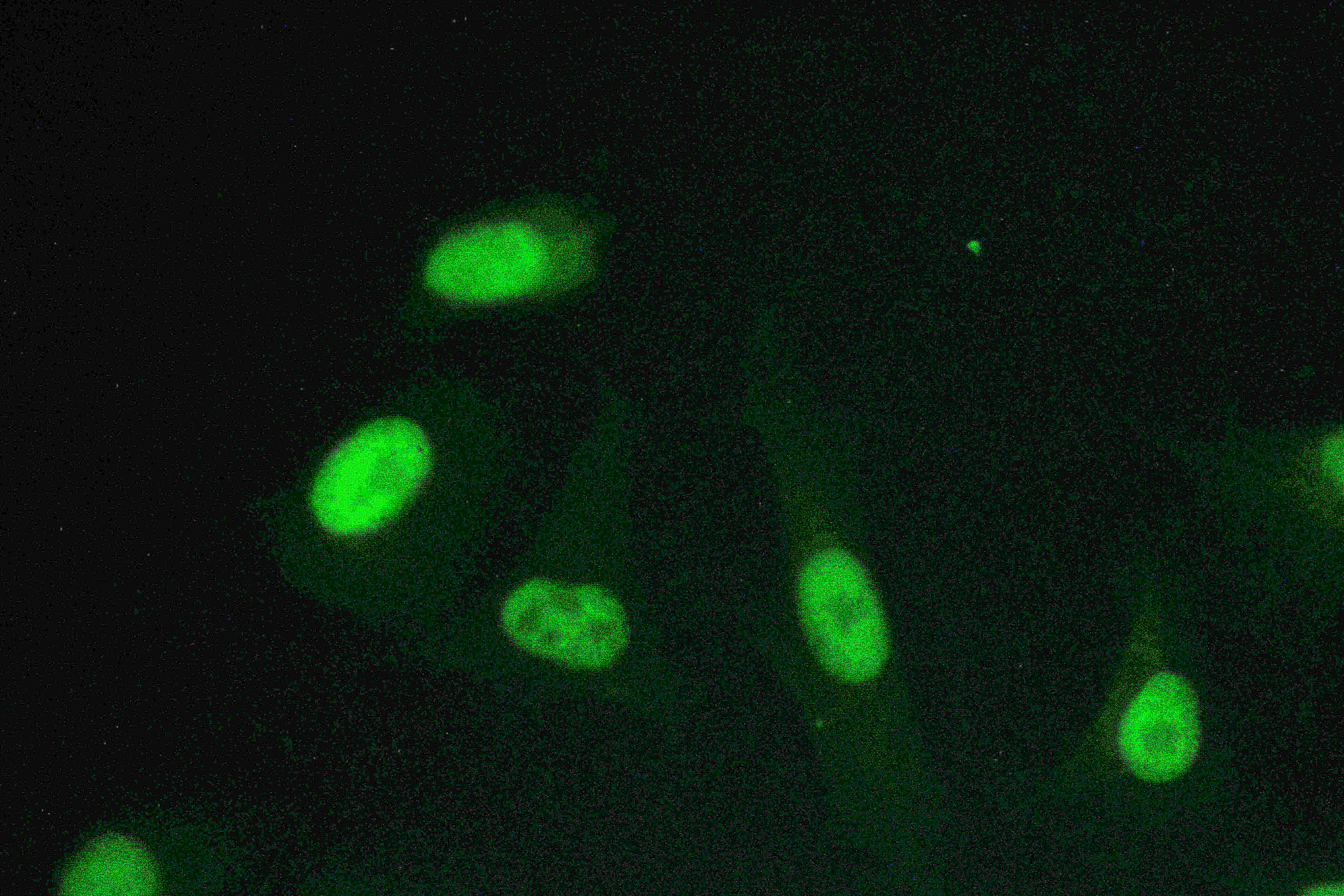
ICC/IF analysis of RPA1 using anti-RPA1 antibody. RPA1 was detected in an immunocytochemical section of A549 cells. The section was incubated with mouse anti-RPA1 Antibody at a dilution of 1:100. Fluoro488-conjugated Anti-mouse IgG Secondary Antibody (green) was used as secondary antibody. The section was counterstained with DAPI (Blue).FC
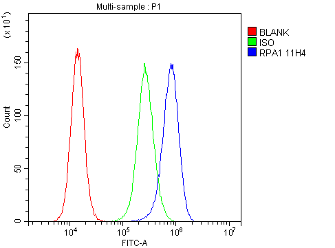
Flow cytometry analysis of A431 cell(1:100) Fluoro 488 conjugated goat anti-mouse IgG(blue) was used as secondary antibody. Isotype control antibody (Green line) was mouse IgG Fluoro 488. Unlabelled sample (Red line).| Product Name | Anti-RPA1 Antibody |
|---|---|
| Antibody Type | Primary Antibodies |
| Immunogen | A synthetic peptide corresponding to a sequence at the C-terminus of human RPA70, different from the related mouse sequence by three amino acids. |
| Clonality | Monoclonal |
|---|---|
| Isotype | IgG2b |
| Host Species | Mouse |
| Tested Applications | FCICC/IFIHCWB |
| WB:1:500-2000 IHC:1:50-400 ICC/IF:1:50-400 FC:1:50-200 |
|
| Species Reactivity | HumanMonkeyMouse |
| Concentration | 1mg/ml |
| Purification | Protein G |
| Gene Symbol | RPA1 |
|---|---|
| Gene Synonyms | HSSB RF-A RP-A REPA1 RPA70 MST075 PFBMFT6 |
| Gene Full Name | replication protein A1 |
| Gene Summary | This gene encodes the largest subunit of the heterotrimeric Replication Protein A (RPA) complex, which binds to single-stranded DNA (ssDNA), forming a nucleoprotein complex that plays an important role in DNA metabolism, being involved in DNA replication, repair, recombination, telomere maintenance, and co-ordinating the cellular response to DNA damage through activation of the ataxia telangiectasia and Rad3-related protein (ATR) kinase. The nucleoprotein complex protects the single-stranded DNA from nucleases, prevents formation of secondary structures that would interfere with repair, and co-ordinates the recruitment and departure of different genome maintenance factors. This subunit contains four oligonucleotide/oligosaccharide-binding (OB) domains, though the majority of ssDNA binding occurs in two of these domains. The heterotrimeric complex has two different modes of ssDNA binding, a low-affinity and high-affinity mode, determined by which ssDNA binding domains are utilized. The different binding modes differ in the length of DNA bound and in the proteins with which it interacts, thereby playing a role in regulating different genomic maintenance pathways. [provided by RefSeq, Sep 2017] |
| Alternative Names | HSSB MST075 REPA1 Replication factor A protein 1 replication protein A1 70kDa RF A RF A protein 1 RP A RP A p70 RPA1 RPA70 |
| Molecular Weight(MW) | 68kDa(Observed MW 72kDa) |
| Cellular Localization | Nucleus |
WB
Western blot analysis of RPA1 using anti-RPA1 antibody. The sample well of each lane was loaded with 30 ug of sample under reducing conditions. Lane 1: HELA whole cell lysates, Lane 2: MCF-7 whole cell lysates, Lane 3: K562 whole cell lysates, Lane 4: COS-7 whole cell lysates, Lane 5: Caco-2 whole cell lysates, Lane 6: A549 whole cell lysates, Lane 7: HEPG2 whole cell lysates, Lane 8: PC-3 whole cell lysates, Lane 9: HEPA1-6 whole cell lysates. After electrophoresis, proteins were transferred to a membrane. Then the membrane was incubated with mouse anti-RPA1 antigen affinity purified monoclonal antibody at a dilution of 1:1000 and probed with a goat anti-mouse IgG-HRP secondary antibody. The signal is developed using ECL Plus Western Blotting Substrate.
IHC
IHC analysis of RPA1 using anti-RPA1 antibody. RPA1 was detected in a paraffin-embedded section of human intestinal cancer tissue. Biotinylated goat anti-mouse IgG was used as secondary antibody. The tissue section was incubated with mouse anti-RPA1 Antibody at a dilution of 1:200 and developed using Strepavidin-Biotin-Complex (SABC) with DAB as the chromogen.
ICC/IF
ICC/IF analysis of RPA1 using anti-RPA1 antibody. RPA1 was detected in an immunocytochemical section of A549 cells. The section was incubated with mouse anti-RPA1 Antibody at a dilution of 1:100. Fluoro488-conjugated Anti-mouse IgG Secondary Antibody (green) was used as secondary antibody. The section was counterstained with DAPI (Blue).
FC
Flow cytometry analysis of A431 cell(1:100) Fluoro 488 conjugated goat anti-mouse IgG(blue) was used as secondary antibody. Isotype control antibody (Green line) was mouse IgG Fluoro 488. Unlabelled sample (Red line).| Application Notes | WB:1:500-2000 IHC:1:50-400 ICC/IF:1:50-400 FC:1:50-200 |
|---|
| Form | Liquid |
|---|---|
| Storage Instructions | 12 months from date of receipt,-20℃ as supplied. |
| Storage Buffer | 500 ug/ml antibody with PBS, 0.02% NaN3, 1 mg/ml BSA and 50% glycerol. |


