WB
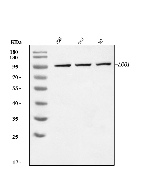
Western blot analysis of AGO1 using anti-AGO1 antibody. The sample well of each lane was loaded with 30 ug of sample under reducing conditions. Lane 1: K562 whole cell lysates, Lane 2: Caco-2 whole cell lysates, Lane 3: 293T whole cell lysates. After electrophoresis, proteins were transferred to a membrane. Then the membrane was incubated with rabbit anti-AGO1 antigen affinity purified polyclonal antibody at a dilution of 1:1000 and probed with a goat anti-rabbit IgG-HRP secondary antibody. The signal is developed using ECL Plus Western Blotting Substrate.ICC/IF
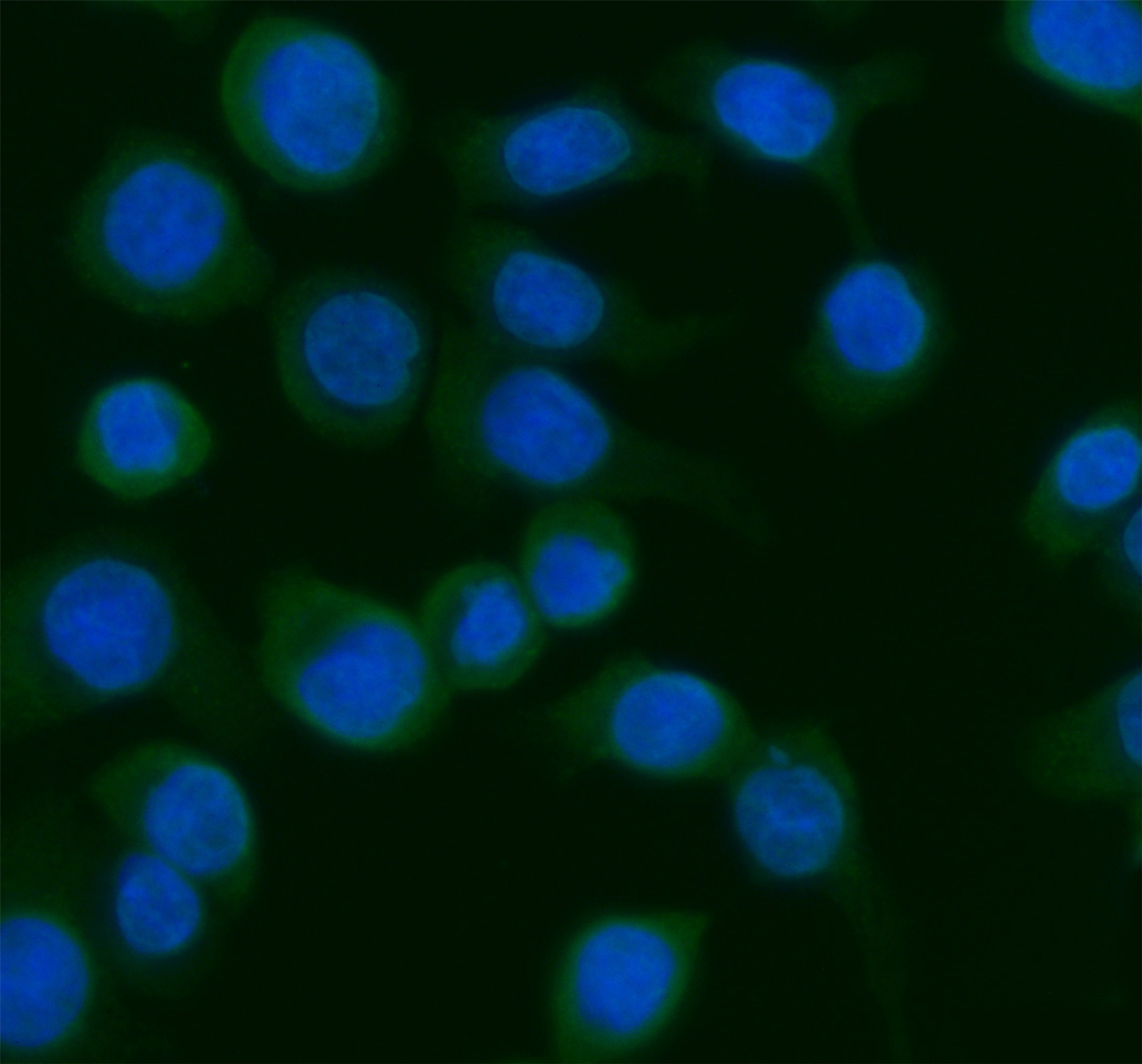
ICC/IF analysis of AGO1 using anti-AGO1 antibody. AGO1 was detected in an immunocytochemical section of Caco-2 cells. The section was incubated with rabbit anti-AGO1 Antibody at a dilution of 1:100. Fluoro488 Conjugated Goat Anti-Rabbit IgG (Green) was used as secondary antibody. The section was counterstained with DAPI(Blue).FC
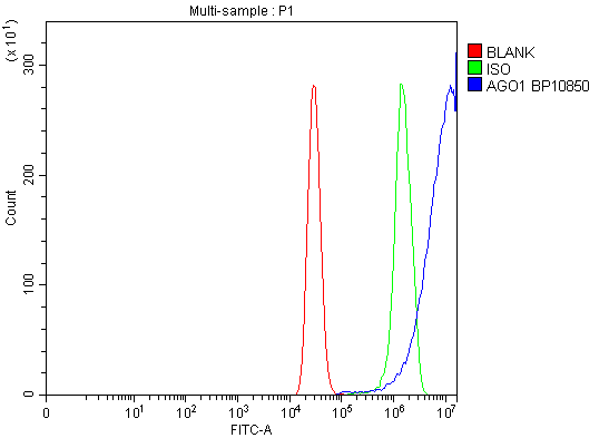
Flow Cytometry analysis of MCF-7 cells using anti-AGO1 antibody. Overlay histogram showing MCF-7 cells stained with anti-AGO1 antibody (Blue line). To facilitate intracellular staining, cells were fixed with 4% paraformaldehyde and permeabilized with permeabilization buffer. The cells were blocked with 10% normal goat serum. And then incubated with rabbit anti-AGO1 Antibody at 1:100 dilution for 30 min at 20°C. Fluoro488 conjugated goat anti-rabbit IgG was used as secondary antibody at 1:100 dilution for 30 minutes at 20°C. Isotype control antibody (Green line) was rabbit IgG at 1:100 dilution used under the same conditions. Unlabelled sample without incubation with primary antibody and secondary antibody (Red line) was used as a blank control.| Product Name | Anti-AGO1 Antibody |
|---|---|
| Antibody Type | Primary Antibodies |
| Immunogen | E.coli-derived human AGO1 recombinant protein (Position: K131-R171). |
| Clonality | Polyclonal |
|---|---|
| Isotype | IgG |
| Host Species | Rabbit |
| Tested Applications | FCICC/IFWB |
| WB:1:500-2000 ICC/IF:1:50-400 FC:1:50-200 |
|
| Species Reactivity | Human |
| Concentration | 1mg/ml |
| Purification | Affinity purified |
| Gene Symbol | AGO1 |
|---|---|
| Gene Synonyms | Q99 EIF2C hAgo1 EIF2C1 GERP95 NEDLBAS |
| Gene Full Name | argonaute RISC component 1 |
| Gene Summary | This gene encodes a member of the argonaute family of proteins, which associate with small RNAs and have important roles in RNA interference (RNAi) and RNA silencing. This protein binds to microRNAs (miRNAs) or small interfering RNAs (siRNAs) and represses translation of mRNAs that are complementary to them. It is also involved in transcriptional gene silencing (TGS) of promoter regions that are complementary to bound short antigene RNAs (agRNAs), as well as in the degradation of miRNA-bound mRNA targets. Alternatively spliced transcript variants encoding different isoforms have been found for this gene. A recent study showed this gene to be an authentic stop codon readthrough target, and that its mRNA could give rise to an additional C-terminally extended isoform by use of an alternative in-frame translation termination codon. [provided by RefSeq, Nov 2015] |
| Alternative Names | AGO1 Argonaute1 eIF 2C 1 EIF2C eIF2C 1 EIF2C1 EIF2C1-Specific GERP95 hAgo1 Protein argonaute 1 Q99 |
| Molecular Weight(MW) | 97kDa(Observed MW 97kDa) |
| Cellular Localization | Cytoplasm, P-body |
WB
Western blot analysis of AGO1 using anti-AGO1 antibody. The sample well of each lane was loaded with 30 ug of sample under reducing conditions. Lane 1: K562 whole cell lysates, Lane 2: Caco-2 whole cell lysates, Lane 3: 293T whole cell lysates. After electrophoresis, proteins were transferred to a membrane. Then the membrane was incubated with rabbit anti-AGO1 antigen affinity purified polyclonal antibody at a dilution of 1:1000 and probed with a goat anti-rabbit IgG-HRP secondary antibody. The signal is developed using ECL Plus Western Blotting Substrate.
ICC/IF
ICC/IF analysis of AGO1 using anti-AGO1 antibody. AGO1 was detected in an immunocytochemical section of Caco-2 cells. The section was incubated with rabbit anti-AGO1 Antibody at a dilution of 1:100. Fluoro488 Conjugated Goat Anti-Rabbit IgG (Green) was used as secondary antibody. The section was counterstained with DAPI(Blue).
FC
Flow Cytometry analysis of MCF-7 cells using anti-AGO1 antibody. Overlay histogram showing MCF-7 cells stained with anti-AGO1 antibody (Blue line). To facilitate intracellular staining, cells were fixed with 4% paraformaldehyde and permeabilized with permeabilization buffer. The cells were blocked with 10% normal goat serum. And then incubated with rabbit anti-AGO1 Antibody at 1:100 dilution for 30 min at 20°C. Fluoro488 conjugated goat anti-rabbit IgG was used as secondary antibody at 1:100 dilution for 30 minutes at 20°C. Isotype control antibody (Green line) was rabbit IgG at 1:100 dilution used under the same conditions. Unlabelled sample without incubation with primary antibody and secondary antibody (Red line) was used as a blank control.| Application Notes | WB:1:500-2000 ICC/IF:1:50-400 FC:1:50-200 |
|---|
| Form | Liquid |
|---|---|
| Storage Instructions | 12 months from date of receipt,-20℃ as supplied. |
| Storage Buffer | 500 ug/ml antibody with PBS, 0.02% NaN3, 1 mg/ml BSA and 50% glycerol. |


