WB
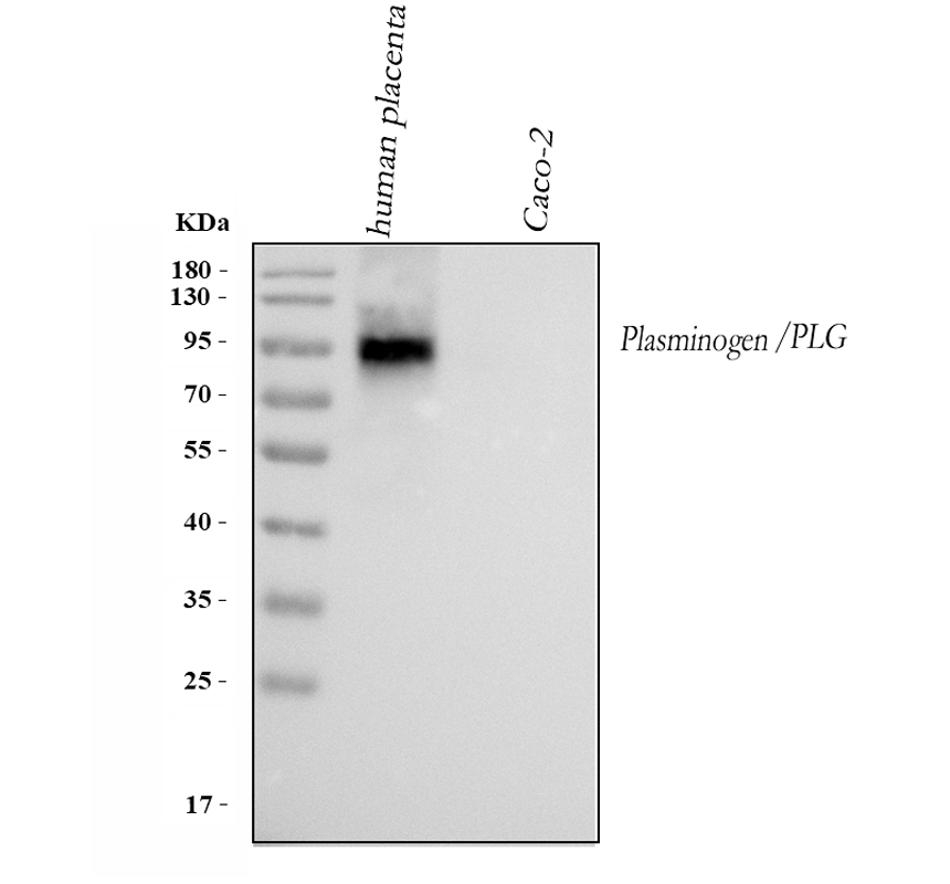
Western blot analysis of Plasminogen/PLG using anti-Plasminogen/PLG antibody. The sample well of each lane was loaded with 30 ug of sample under reducing conditions. Lane 1: human placenta tissue lysates, Lane 2: human Caco-2 whole cell lysates. After electrophoresis, proteins were transferred to a membrane. Then the membrane was incubated with rabbit anti-Plasminogen/PLG antigen affinity purified polyclonal antibody at a dilution of 1:1000 and probed with a goat anti-rabbit IgG-HRP secondary antibody. The signal is developed using ECL Plus Western Blotting Substrate.IHC
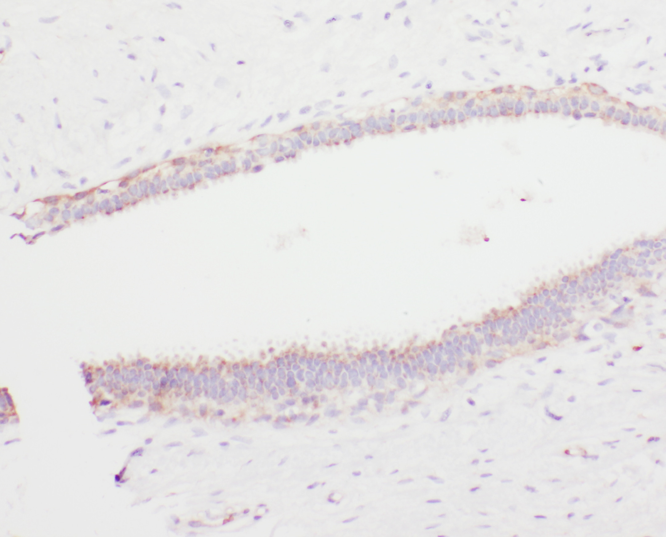
IHC analysis of Plasminogen/PLG using anti-Plasminogen/PLG antibody. Plasminogen/PLG was detected in a paraffin-embedded section of human mammary cancer tissue. Biotinylated goat anti-rabbit IgG was used as secondary antibody. The tissue section was incubated with rabbit anti-Plasminogen/PLG Antibody at a dilution of 1:200 and developed using Strepavidin-Biotin-Complex (SABC) with DAB as the chromogen.| Product Name | Anti-Plasminogen/PLG Antibody |
|---|---|
| Antibody Type | Primary Antibodies |
| Immunogen | E. coli-derived human Angiostatin K1-3 recombinant protein(Position: C103-C352). |
| Clonality | Polyclonal |
|---|---|
| Isotype | IgG |
| Host Species | Rabbit |
| Tested Applications | IHCWB |
| WB:1:500-2000 IHC:1:50-400 |
|
| Species Reactivity | Human |
| Concentration | 1mg/ml |
| Purification | Affinity purified |
| Gene Symbol | PLG |
|---|---|
| Gene Synonyms | HAE4 |
| Gene Full Name | plasminogen |
| Gene Summary | The plasminogen protein encoded by this gene is a serine protease that circulates in blood plasma as an inactive zymogen and is converted to the active protease, plasmin, by several plasminogen activators such as tissue plasminogen activator (tPA), urokinase plasminogen activator (uPA), kallikrein, and factor XII (Hageman factor). The conversion of plasminogen to plasmin involves the cleavage of the peptide bond between Arg-561 and Val-562. Plasmin cleavage also releases the angiostatin protein which inhibits angiogenesis. Plasmin degrades many blood plasma proteins, including fibrin-containing blood clots. As a serine protease, plasmin cleaves many products in addition to fibrin such as fibronectin, thrombospondin, laminin, and von Willebrand factor. Plasmin is inactivated by proteins such as alpha-2-macroglobulin and alpha-2-antiplasmin in addition to inhibitors of the various plasminogen activators. Plasminogen also interacts with plasminogen receptors which results in the retention of plasmin on cell surfaces and in plasmin-induced cell signaling. The localization of plasminogen on cell surfaces plays a role in the degradation of extracellular matrices, cell migration, inflamation, wound healing, oncogenesis, metastasis, myogenesis, muscle regeneration, neurite outgrowth, and fibrinolysis. This protein may also play a role in acute respiratory distress syndrome (ARDS) which, in part, is caused by enhanced clot formation and the suppression of fibrinolysis. Compared to other mammals, the cluster of plasminogen-like genes to which this gene belongs has been rearranged in catarrhine primates. [provided by RefSeq, May 2020] |
| Alternative Names | Angiostatin plasminogen PLG |
| Molecular Weight(MW) | 91kDa(Observed MW 90kDa) |
| Cellular Localization | Secreted |
WB
Western blot analysis of Plasminogen/PLG using anti-Plasminogen/PLG antibody. The sample well of each lane was loaded with 30 ug of sample under reducing conditions. Lane 1: human placenta tissue lysates, Lane 2: human Caco-2 whole cell lysates. After electrophoresis, proteins were transferred to a membrane. Then the membrane was incubated with rabbit anti-Plasminogen/PLG antigen affinity purified polyclonal antibody at a dilution of 1:1000 and probed with a goat anti-rabbit IgG-HRP secondary antibody. The signal is developed using ECL Plus Western Blotting Substrate.
IHC
IHC analysis of Plasminogen/PLG using anti-Plasminogen/PLG antibody. Plasminogen/PLG was detected in a paraffin-embedded section of human mammary cancer tissue. Biotinylated goat anti-rabbit IgG was used as secondary antibody. The tissue section was incubated with rabbit anti-Plasminogen/PLG Antibody at a dilution of 1:200 and developed using Strepavidin-Biotin-Complex (SABC) with DAB as the chromogen.| Application Notes | WB:1:500-2000 IHC:1:50-400 |
|---|
| Form | Liquid |
|---|---|
| Storage Instructions | 12 months from date of receipt,-20℃ as supplied. |
| Storage Buffer | 500 ug/ml antibody with PBS, 0.02% NaN3, 1 mg/ml BSA and 50% glycerol. |


