WB
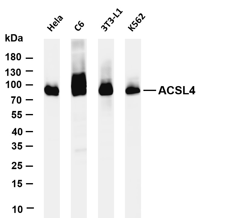
Various whole cell lysates were separated by 4-20% SDS-PAGE, and the membrane was blotted with anti-ACSL4 antibody. The HRP-conjugated Goat anti-Rabbit IgG(H + L) antibody was used to detect the antibody. Lane 1: Hela Lane 2: C6 Lane 3: 3T3-L1 Lane 4: K562.WB
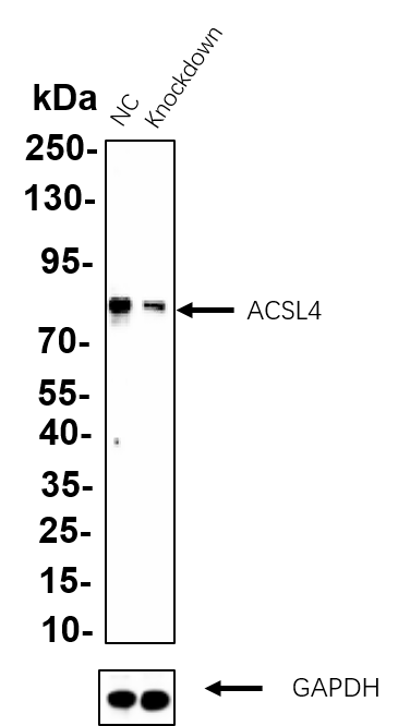
Western blot analysis of lysates from Hela WT and knockdown cell. Cell lysates were separated by 4-20% SDS-PAGE, and the membrane was blotted with FACL4 rabbit mAb. The HRP-conjugated Goat anti-Rabbit IgG (H + L) antibody was used to detect the antibody.IHC
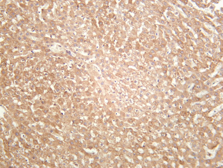
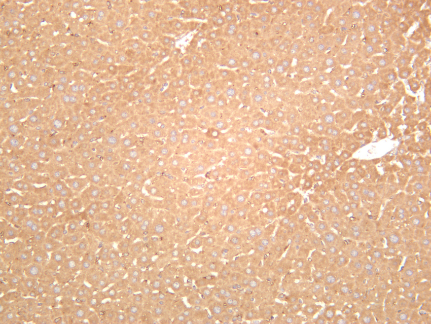
Rat liver was stained with anti-ACSL4 rabbit antibody.IHC
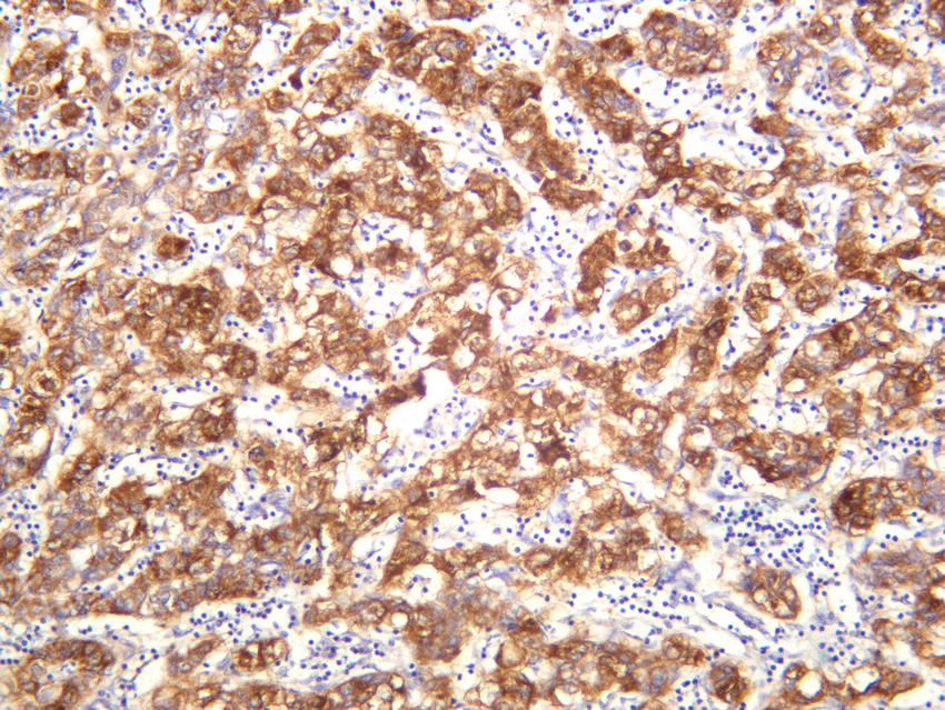
Human hepatocellular carcinoma was stained with anti-ACSL4 rabbit antibody.IHC
Mouse liver was stained with anti-ACSL4 rabbit antibody.ICC/IF
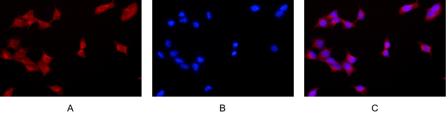
Immunofluorescence analysis of HEK293. Picture A: ACSL4 antibody (red). Picture B: DAPI (blue). Picture C: Merge of A+B.| Product Name | ACSL4 Rabbit mAb |
|---|---|
| Antibody Type | Primary Antibodies |
| Clonality | monoclonal |
|---|---|
| Isotype | IgG,K |
| Host Species | Rabbit |
| Tested Applications | ICC/IFIHCWB |
| WB:1:1000-1:5000 IHC:1:200-1:2000 ICC/IF:1:200-1:1000 |
|
| Species Reactivity | HumanMouseRat |
| Concentration | 1mg/ml |
| Purification | Protein A |
| Gene Symbol | ACSL4 |
|---|---|
| Gene Synonyms | ACS4 FACL4 LACS4 MRX63 MRX68 XLID63 |
| Gene Full Name | acyl-CoA synthetase long chain family member 4 |
| Gene Summary | The protein encoded by this gene is an isozyme of the long-chain fatty-acid-coenzyme A ligase family. Although differing in substrate specificity, subcellular localization, and tissue distribution, all isozymes of this family convert free long-chain fatty acids into fatty acyl-CoA esters, and thereby play a key role in lipid biosynthesis and fatty acid degradation. This isozyme preferentially utilizes arachidonate as substrate. The absence of this enzyme may contribute to the cognitive disability or Alport syndrome. Alternative splicing of this gene generates multiple transcript variants. [provided by RefSeq, Jan 2016] |
| Alternative Names | Long-chain-fatty-acid--CoA ligase 4 Long-chain acyl-CoA synthetase 4 LACS 4 |
| Molecular Weight(MW) | 78kDa(Observed MW 78kDa) |
| Cellular Localization | Membrane |
WB
Various whole cell lysates were separated by 4-20% SDS-PAGE, and the membrane was blotted with anti-ACSL4 antibody. The HRP-conjugated Goat anti-Rabbit IgG(H + L) antibody was used to detect the antibody. Lane 1: Hela Lane 2: C6 Lane 3: 3T3-L1 Lane 4: K562.
WB
Western blot analysis of lysates from Hela WT and knockdown cell. Cell lysates were separated by 4-20% SDS-PAGE, and the membrane was blotted with FACL4 rabbit mAb. The HRP-conjugated Goat anti-Rabbit IgG (H + L) antibody was used to detect the antibody.
IHC
Rat liver was stained with anti-ACSL4 rabbit antibody.
IHC
Human hepatocellular carcinoma was stained with anti-ACSL4 rabbit antibody.
IHC
Mouse liver was stained with anti-ACSL4 rabbit antibody.
ICC/IF
Immunofluorescence analysis of HEK293. Picture A: ACSL4 antibody (red). Picture B: DAPI (blue). Picture C: Merge of A+B.| Application Notes | WB:1:1000-1:5000 IHC:1:200-1:2000 ICC/IF:1:200-1:1000 |
|---|
| Form | Liquid |
|---|---|
| Storage Instructions | -15°C to -25°C/1 year(Do not lower than -25°C) |
| Storage Buffer | PBS, 50% glycerol, 0.05% Proclin 300, 0.05%BSA |


