WB
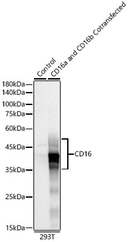
Western blot analysis of lysates from wild type (WT) and 293F cells cotransfected with CD16a and CD16b,using CD16 Rabbit mAb at 1:2000 dilution.Secondary antibody: HRP-conjugated Goat anti-Rabbit IgG (H+L) at 1:10000 dilution.Lysates/proteins: 25μg per lane.Blocking buffer: 3% nonfat dry milk in TBST.Detection: ECL Basic Kit.Exposure time: 10s.WB
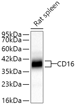
Western blot analysis of lysates from Rat spleen using CD16 Rabbit mAb at 1:1000 dilutionincubated overnight at 4℃.Secondary antibody: HRP-conjugated Goat anti-Rabbit IgG (H+L) at 1:10000 dilution.Lysates/proteins: 25 μg per lane.Blocking buffer: 3% nonfat dry milk in TBST.Detection: ECL Basic Kit.Exposure time: 30 s.IHC
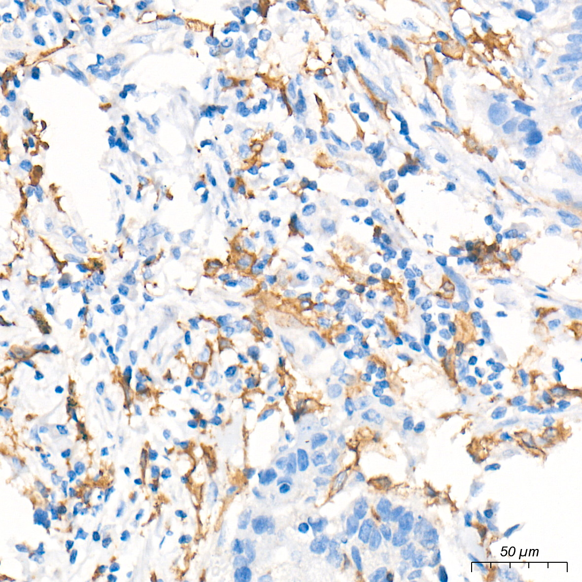
Immunohistochemistry analysis of paraffinembedded Human liver tissue using CD16Rabbit mAb at a dilution of 1:5000(40x lens). High pressure antigen retrievalperformed with 0.01M Tris-EDTA Buffer (pH9.0) prior to IHC staining.ICC/IF
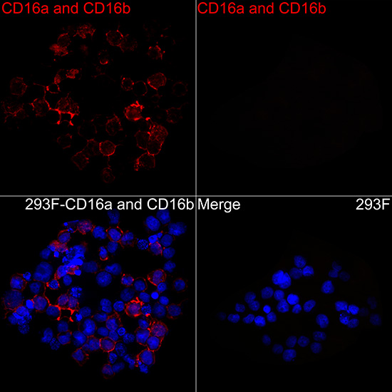
Immunofluorescence analysis of 293F cellstransfected with CD16a and CD16b usingCD16 Rabbit mAb at dilution of1:100 (40x lens). Secondary antibody: Cy3-conjugated Goat anti-Rabbit IgG (H+L) at 1:500 dilution. Blue: DAPI fornuclear staining.| Product Name | CD16 Rabbit mAb |
|---|---|
| Antibody Type | Primary Antibodies |
| Immunogen | Recombinant protein (or fragment).This information is considered to be commercially sensitive. |
| Clonality | Monoclonal |
|---|---|
| Isotype | IgG |
| Host Species | Rabbit |
| Tested Applications | ICC/IFIHCWB |
| WB:1:1000-1:3000 IHC:1:2000-1:8000 ICC/IF:1:50-1:200 |
|
| Species Reactivity | HumanRat |
| Concentration | 1mg/ml |
| Purification | Affinity purified |
| Gene Symbol | FCGR3A |
|---|---|
| Gene Synonyms | FCG3 CD16A FCGR3 IGFR3 IMD20 CD16-II FCRIIIA FcGRIIIA |
| Gene Full Name | Fc gamma receptor IIIa |
| Gene Summary | This gene encodes a receptor for the Fc portion of immunoglobulin G, and it is involved in the removal of antigen-antibody complexes from the circulation, as well as other responses, including antibody dependent cellular mediated cytotoxicity and antibody dependent enhancement of virus infections. This gene (FCGR3A) is highly similar to another nearby gene (FCGR3B) located on chromosome 1. The receptor encoded by this gene is expressed on natural killer (NK) cells as an integral membrane glycoprotein anchored through a transmembrane peptide, whereas FCGR3B is expressed on polymorphonuclear neutrophils (PMN) where the receptor is anchored through a phosphatidylinositol (PI) linkage. Mutations in this gene are associated with immunodeficiency 20, and have been linked to susceptibility to recurrent viral infections, susceptibility to systemic lupus erythematosus, and alloimmune neonatal neutropenia. Alternatively spliced transcript variants encoding different isoforms have been found for this gene. [provided by RefSeq, Aug 2020] |
| Alternative Names | CD16 FCG3 CD16A FCGR3 IGFR3 IMD20 FCR-10 FCRIII CD16-II FCGRIII FCRIIIA FcGRIIIA |
| Molecular Weight(MW) | 29/26kDa(Observed band size:40-60kDa) |
| Cellular Localization | Secreted , Cell membrane, Single-pass type I membrane protein(FCGR3A) ;Secreted , Cell membrane, Lipid-anchor, GPI-anchor(FCGR3B). |
WB
Western blot analysis of lysates from wild type (WT) and 293F cells cotransfected with CD16a and CD16b,using CD16 Rabbit mAb at 1:2000 dilution.Secondary antibody: HRP-conjugated Goat anti-Rabbit IgG (H+L) at 1:10000 dilution.Lysates/proteins: 25μg per lane.Blocking buffer: 3% nonfat dry milk in TBST.Detection: ECL Basic Kit.Exposure time: 10s.
WB
Western blot analysis of lysates from Rat spleen using CD16 Rabbit mAb at 1:1000 dilutionincubated overnight at 4℃.Secondary antibody: HRP-conjugated Goat anti-Rabbit IgG (H+L) at 1:10000 dilution.Lysates/proteins: 25 μg per lane.Blocking buffer: 3% nonfat dry milk in TBST.Detection: ECL Basic Kit.Exposure time: 30 s.
IHC
Immunohistochemistry analysis of paraffinembedded Human liver tissue using CD16Rabbit mAb at a dilution of 1:5000(40x lens). High pressure antigen retrievalperformed with 0.01M Tris-EDTA Buffer (pH9.0) prior to IHC staining.
ICC/IF
Immunofluorescence analysis of 293F cellstransfected with CD16a and CD16b usingCD16 Rabbit mAb at dilution of1:100 (40x lens). Secondary antibody: Cy3-conjugated Goat anti-Rabbit IgG (H+L) at 1:500 dilution. Blue: DAPI fornuclear staining.| Application Notes | WB:1:1000-1:3000 IHC:1:2000-1:8000 ICC/IF:1:50-1:200 |
|---|
| Form | Liquid |
|---|---|
| Storage Instructions | Store at -20℃. Avoid freeze / thaw cycles. |
| Storage Buffer | PBS containing 50% glycerol and 0.05% BSA, preserved with proclin300 or sodium azide (as specified on the Certificate of Analysis), pH 7.3. |


