WB
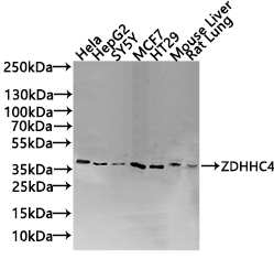
Western Blot, Positive WB detected in: Hela whole cell lysate(30µg), HepG2 whole cell lysate(30µg), SY5Y whole cell lysate(30µg), MCF7 whole cell lysate(30µg), HT29 whole cell lysate(30µg),Mouse Liver tissue lysate(30µg),Rat Lung tissue lysate(30µg) All lanes: ZDHHC4 antibody at 1:1000, Secondary, Goat polyclonal to rabbit IgG at 1/20000 dilution, Exposure time: 120s.IHC
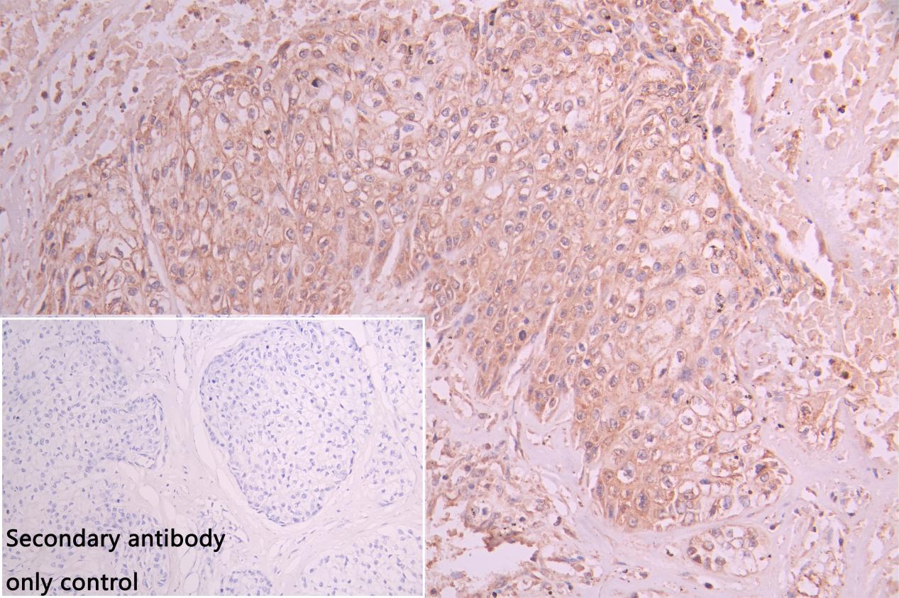
IHC image of ZDHHC4 Antibody diluted at 1:50 and staining in paraffin-embedded human pancreatic tissue performed on a Leica BondTM system. After dewaxing and hydration, antigen retrieval was mediated by high pressure in a citrate buffer (pH 6.0). Section was blocked with 10% normal goat serum 30min at RT. Then primary antibody (1% BSA) was incubated at 4°C overnight. The primary is detected by a Goat anti-rabbit polymer IgG labeled by HRP and visualized using 0.05% DAB. Secondary antibody only control: uses 1% BSA instead of primary antibody.| Product Name | ZDHHC4 Antibody |
|---|---|
| Antibody Type | Primary Antibodies |
| Immunogen | Recombinant Human Probable palmitoyltransferase ZDHHC4 protein (277-344AA) |
| Clonality | Polyclonal |
|---|---|
| Isotype | IgG |
| Host Species | Rabbit |
| Tested Applications | IHCWB |
| WB:1:500-1:3000 IHC:1:20-1:200 |
|
| Species Reactivity | HumanMouseRat |
| Concentration | 1mg/ml |
| Purification | Protein G |
| Gene Symbol | ZDHHC4 |
|---|---|
| Gene Synonyms | ZNF374 |
| Gene Full Name | zDHHC palmitoyltransferase 4 |
| Gene Summary | Enables palmitoyltransferase activity. Involved in positive regulation of innate immune response and protein localization to plasma membrane. Located in Golgi apparatus and endoplasmic reticulum. [provided by Alliance of Genome Resources, Jul 2025] |
| Molecular Weight(MW) | 40kDa(Observed MW 40kDa) |
| Cellular Localization | Endoplasmic reticulum membrane; Multi-pass membrane protein. Golgi apparatus membrane; Multi-pass membrane protein. Cell membrane; Multi-pass membrane protein. |
WB
Western Blot, Positive WB detected in: Hela whole cell lysate(30µg), HepG2 whole cell lysate(30µg), SY5Y whole cell lysate(30µg), MCF7 whole cell lysate(30µg), HT29 whole cell lysate(30µg),Mouse Liver tissue lysate(30µg),Rat Lung tissue lysate(30µg) All lanes: ZDHHC4 antibody at 1:1000, Secondary, Goat polyclonal to rabbit IgG at 1/20000 dilution, Exposure time: 120s.
IHC
IHC image of ZDHHC4 Antibody diluted at 1:50 and staining in paraffin-embedded human pancreatic tissue performed on a Leica BondTM system. After dewaxing and hydration, antigen retrieval was mediated by high pressure in a citrate buffer (pH 6.0). Section was blocked with 10% normal goat serum 30min at RT. Then primary antibody (1% BSA) was incubated at 4°C overnight. The primary is detected by a Goat anti-rabbit polymer IgG labeled by HRP and visualized using 0.05% DAB. Secondary antibody only control: uses 1% BSA instead of primary antibody.| Application Notes | WB:1:500-1:3000 IHC:1:20-1:200 |
|---|
| Form | Liquid |
|---|---|
| Storage Instructions | Upon receipt, store at -20°C or -80°C. Avoid repeated freeze. |
| Storage Buffer | Preservative: 0.03% Proclin 300 Constituents: 50% Glycerol, 0.01M PBS, pH 7.4 |


