WB
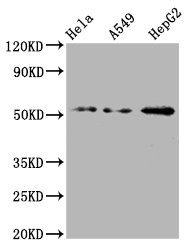
Western Blot, Positive WB detected in: Hela whole cell lysate, A549 whole cell lysate, HepG2 whole cell lysate, All lanes: CD47 antibody at 1:2000, Secondary, Goat polyclonal to rabbit IgG at 1/50000 dilution.IHC
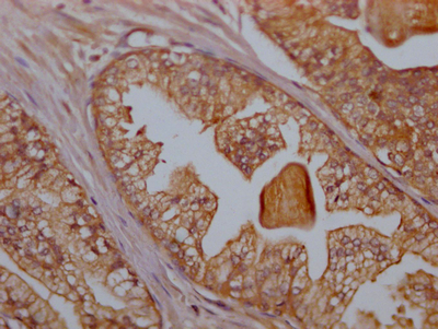
IHC image of CD47 Recombinant Monoclonal Antibody diluted at 1:100 and staining in paraffin-embedded human prostate cancer performed on a Leica BondTM system. After dewaxing and hydration, antigen retrieval was mediated by high pressure in a citrate buffer (pH 6.0). Section was blocked with 10% normal goat serum 30min at RT. Then primary antibody (1% BSA) was incubated at 4℃ overnight. The primary is detected by a Goat anti-rabbit IgG polymer labeled by HRP and visualized using 0.05% DAB.ICC/IF
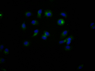
Immunofluorescence staining of MCF7 Cells with CD47 Recombinant Monoclonal Antibody at 1:50, counter-stained with DAPI. The cells were fixed in 4% formaldehyde, permeated by 0.2% TritonX-100, and blocked in 10% normal Goat Serum. The cells were then incubated with the antibody overnight at 4℃. Nuclear DNA was labeled in blue with DAPI. The secondary antibody was FITC-conjugated AffiniPure Goat Anti-Rabbit IgG (H+L).| Product Name | CD47 Recombinant Monoclonal Antibody |
|---|---|
| Antibody Type | Primary Antibodies |
| Immunogen | A synthesized peptide derived from human CD47 |
| Clonality | Monoclonal |
|---|---|
| Isotype | IgG |
| Host Species | Rabbit |
| Tested Applications | ICC/IFIHCWB |
| WB:1:500-1:2000 IHC:1:50-1:200 ICC/IF:1:20-1:200 |
|
| Species Reactivity | Human |
| Concentration | 1mg/ml |
| Purification | Affinity purified |
| Gene Symbol | CD47 |
|---|---|
| Gene Synonyms | IAP OA3 MER6 |
| Gene Full Name | CD47 molecule |
| Gene Summary | This gene encodes a membrane protein, which is involved in the increase in intracellular calcium concentration that occurs upon cell adhesion to extracellular matrix. The encoded protein is also a receptor for the C-terminal cell binding domain of thrombospondin, and it may play a role in membrane transport and signal transduction. This gene has broad tissue distribution, and is reduced in expression on Rh erythrocytes. Alternatively spliced transcript variants have been found for this gene. [provided by RefSeq, Jul 2010] |
| Molecular Weight(MW) | 36/32/34kDa(Observed MW 55kDa) |
| Cellular Localization | Cell membrane; Multi-pass membrane protein. |
WB
Western Blot, Positive WB detected in: Hela whole cell lysate, A549 whole cell lysate, HepG2 whole cell lysate, All lanes: CD47 antibody at 1:2000, Secondary, Goat polyclonal to rabbit IgG at 1/50000 dilution.
IHC
IHC image of CD47 Recombinant Monoclonal Antibody diluted at 1:100 and staining in paraffin-embedded human prostate cancer performed on a Leica BondTM system. After dewaxing and hydration, antigen retrieval was mediated by high pressure in a citrate buffer (pH 6.0). Section was blocked with 10% normal goat serum 30min at RT. Then primary antibody (1% BSA) was incubated at 4℃ overnight. The primary is detected by a Goat anti-rabbit IgG polymer labeled by HRP and visualized using 0.05% DAB.
ICC/IF
Immunofluorescence staining of MCF7 Cells with CD47 Recombinant Monoclonal Antibody at 1:50, counter-stained with DAPI. The cells were fixed in 4% formaldehyde, permeated by 0.2% TritonX-100, and blocked in 10% normal Goat Serum. The cells were then incubated with the antibody overnight at 4℃. Nuclear DNA was labeled in blue with DAPI. The secondary antibody was FITC-conjugated AffiniPure Goat Anti-Rabbit IgG (H+L).| Application Notes | WB:1:500-1:2000 IHC:1:50-1:200 ICC/IF:1:20-1:200 |
|---|
| Form | Liquid |
|---|---|
| Storage Instructions | Upon receipt, store at -20°C or -80°C. Avoid repeated freeze. |
| Storage Buffer | Rabbit IgG in 10mM phosphate buffered saline , pH 7.4, 150mM sodium chloride, 0.05% BSA, 0.02% sodium azide and 50% glycerol. |


