WB
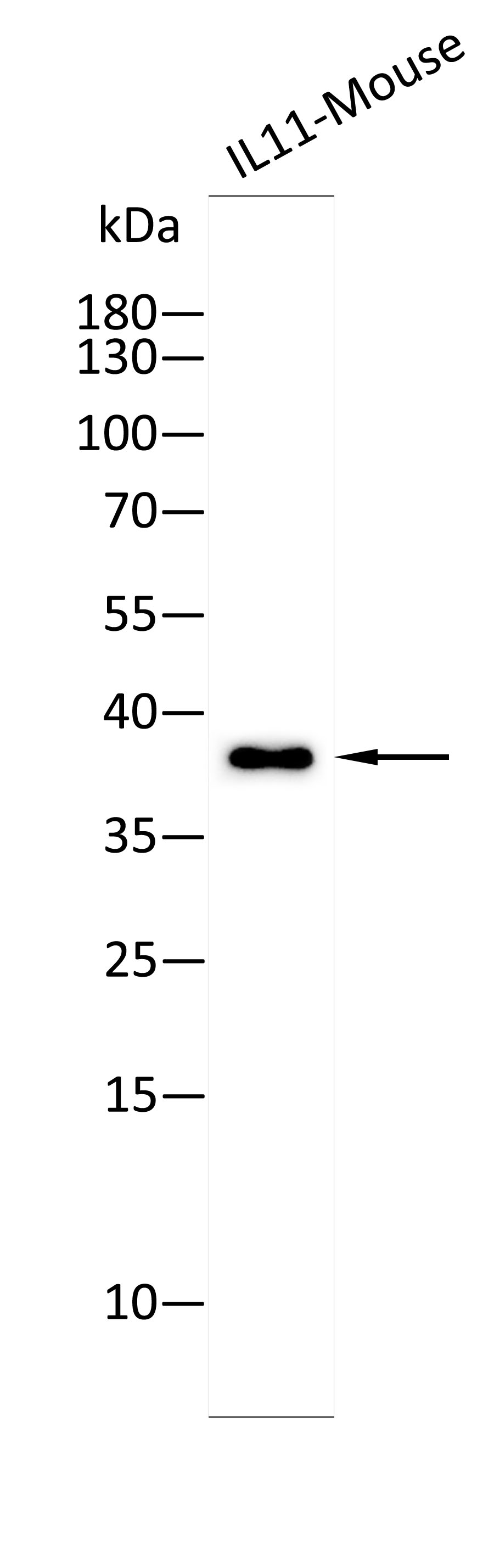
Western blot analysis using Anti-IL11-Mouse antibody against Recombinant IL11-Mouse protein. 15% SDS-PAGE gel.Sample loading: 10ng /lane. Transfer the proteins onto a PVDF membrane (OM790002), and block it with TBST (OM750016) plus skimmed milk powder(OM750084) for one hour. Dilute the primary antibody with the antibody diluent (OM750012) at a ratio of 1:5000, and incubate it overnight at 4°C. Wash the membrane three times with TBST (OM750016), 5 minutes each time. At room temperature, dilute the secondary antibody, Goat Anti-Rabbit IgG(H&L)-HRP (OM643487), at a ratio of 1:10000 and incubate for one hour. Wash the membrane three times with TBST (OM750016) again, 5 minutes each time. Use ECL (OM625701) for luminescence.staining time: 30S.WB
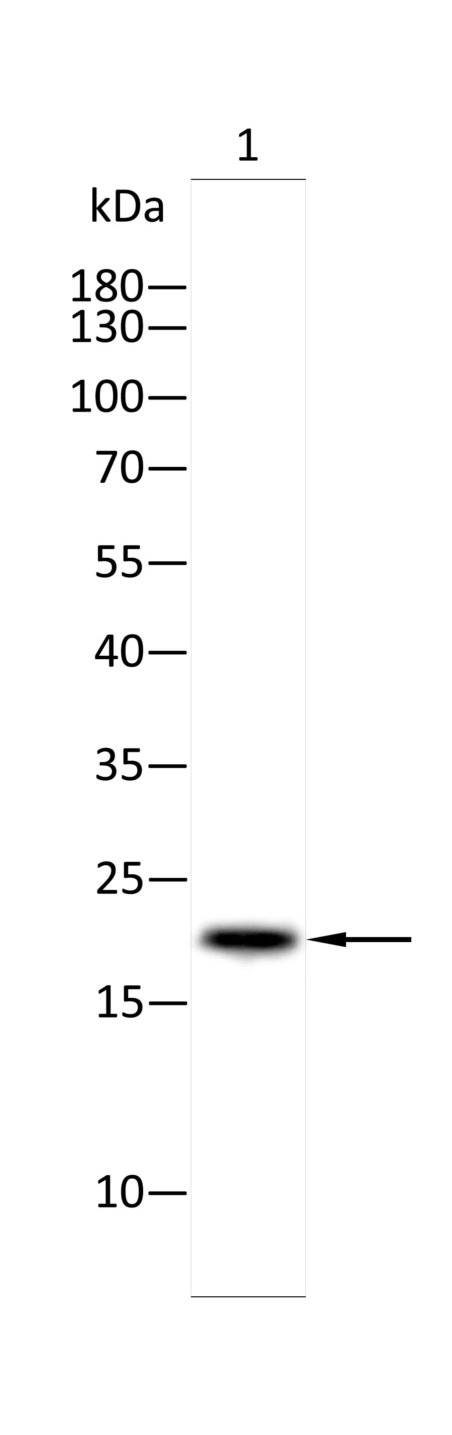
Western blot analysis using Anti-IL11-Mouse antibody against MFC(1) cell. 15% SDS-PAGE gel.Sample loading: 20ug /lane. Transfer the proteins onto a PVDF membrane (OM790002), and block it with TBST (OM750016) plus skimmed milk powder(OM750084) for one hour. Dilute the primary antibody with the antibody diluent (OM750012) at a ratio of 1:2000, and incubate it overnight at 4°C. Wash the membrane three times with TBST (OM750016), 5 minutes each time. At room temperature, dilute the secondary antibody, Goat Anti-Rabbit IgG(H&L)-HRP (OM643487), at a ratio of 1:10000 and incubate for one hour. Wash the membrane three times with TBST (OM750016) again, 5 minutes each time. Use ECL (OM625701) for luminescence.staining time: 60S.WB
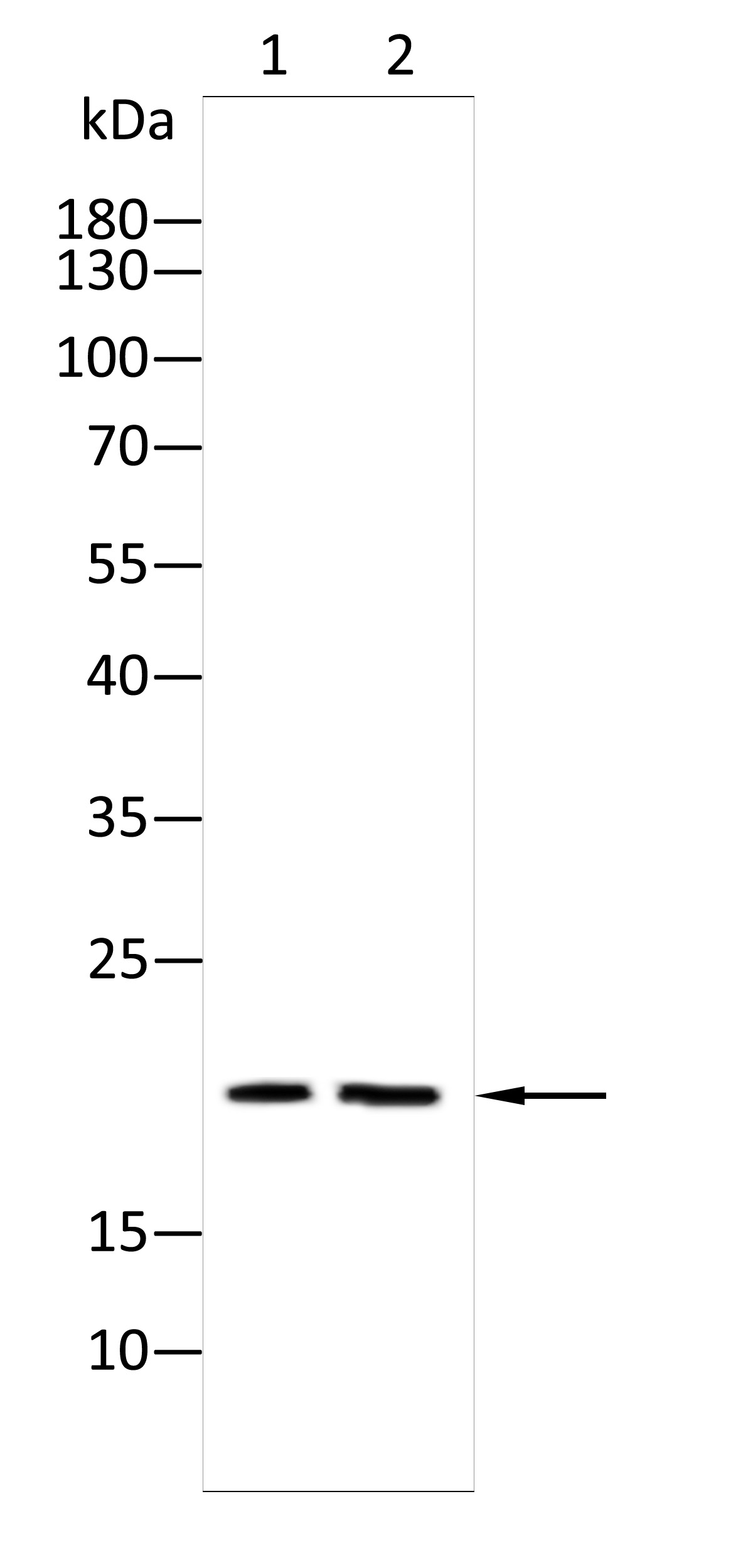
Western blot analysis using IL11 antibody against Nih3t3(1) cell,Mouse brain tissue(2) . 12% SDS-PAGE gel.Sample loading: 20μg /lane. Transfer the proteins onto a PVDF membrane (OM790003), and block it with TBST (OM750016) plus skimmed milk powder(OM750084) for one hour. Dilute the primary antibody with the antibody diluent (OM750012) at a ratio of 1:1000, and incubate it overnight at 4°C. Wash the membrane three times with TBST (OM750016), 5 minutes each time. At room temperature, dilute the secondary antibody, Goat Anti-Rabbit IgG(H&L)-HRP (OM643487), at a ratio of 1:20000 and incubate for one hour. Wash the membrane three times with TBST (OM750016) again, 5 minutes each time. Use ECL (OM625701) for luminescence.staining time: 60S.ELISA
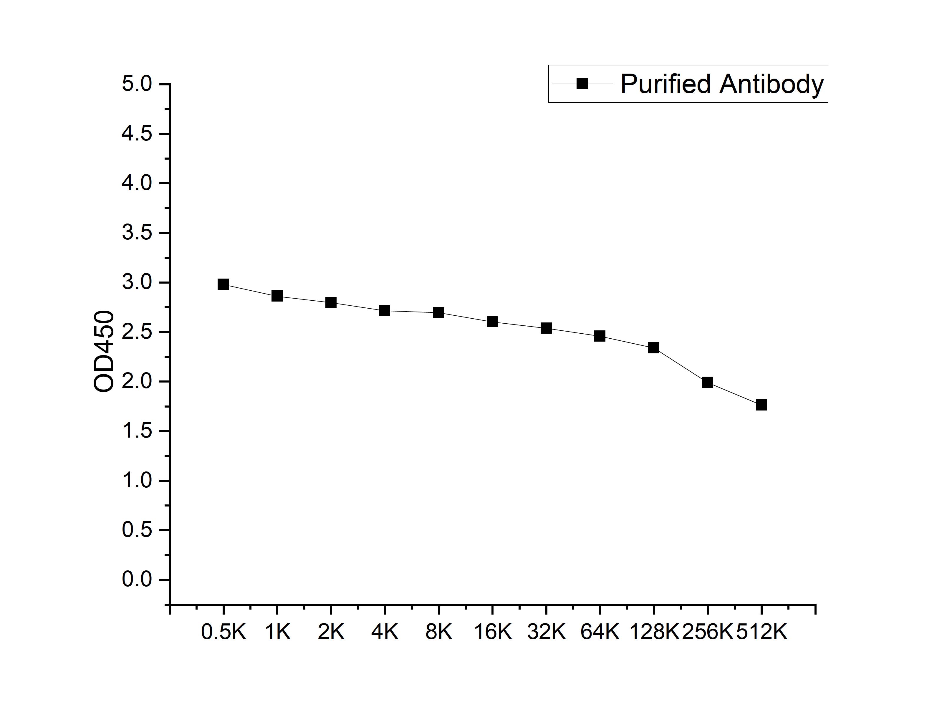
Microtiter plate wells were coated with 100 ng/well coating antigen and incubated with serial dilutions of the antibody. Following HRP-conjugated secondary antibody and substrate development, OD450 was measured. The absorbance decreased progressively with increasing antibody dilution, yielding a sigmoidal titration curve that demonstrates the high antigen-binding activity of the polyclonal antibody.ICC/IF
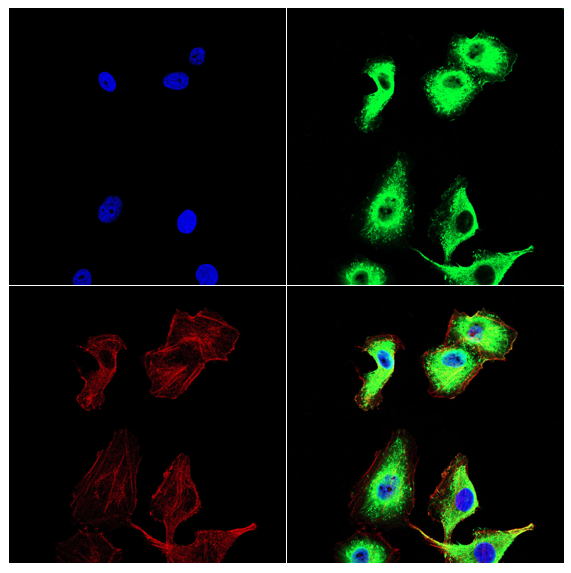
Immunofluorescence analysis of HUVECcells using IL11 antibody (green). Blue: DAPI fluorescent DNA dye. Red: Actin filaments have been labeled with Omnimabs® 594-Phalloidin.Cells are fixed in 4% paraformaldehyde at room temperature for 15 minutes. Then, they are permeabilized with a PBS (OM750003) solution containing 0.1% Triton X-100(OM750021) at room temperature for 15 minutes. Subsequently, the cells are blocked with 10% non - immune goat serum(OM760028) at room temperature for 1 hour.The cells are incubated overnight at 4°C with the primary antibody diluted 1:100 in PBS. The secondary antibody, Omnimabs® 488 Goat Ant-Rabbit IgG(H&L) (Green,OM643486), is diluted at a ratio of 1:400 and incubated with the cells for 1 hour.Nuclear DNA is labeled with DAPI (Blue,OM643160). F-actin is stained with Omnimabs® 594-Phalloidin (Red,OM750007) diluted 1:100 for 30 minutes.| Product Name | Anti-IL11 antibody |
|---|---|
| Antibody Type | Primary Antibodies |
| Immunogen | Recombinant Mouse IL11 protein |
| Clonality | polyclonal |
|---|---|
| Isotype | IgG |
| Host Species | Rabbit |
| Tested Applications | ELISAICC/IFWB |
| WB:1:1000-1:2000 ICC/IF:1:100-1:500 |
|
| Species Reactivity | Mouse |
| Concentration | 1mg/ml |
| Purification | Affinity purified |
| Gene Symbol | IL11 |
|---|---|
| Gene Synonyms | AGIF IL-11 |
| Gene Full Name | interleukin 11 |
| Gene Summary | The protein encoded by this gene is a member of the gp130 family of cytokines. These cytokines drive the assembly of multisubunit receptor complexes, all of which contain at least one molecule of the transmembrane signaling receptor IL6ST (gp130). This cytokine is shown to stimulate the T-cell-dependent development of immunoglobulin-producing B cells. It is also found to support the proliferation of hematopoietic stem cells and megakaryocyte progenitor cells. Alternatively spliced transcript variants encoding distinct isoforms have been found for this gene. [provided by RefSeq, Jun 2012] |
| Alternative Names | adipogenesis inhibitory factor il 11 IL-11ra IL10RA IL11 inhibitor ILN Interleukin Interleukin11 MGC2146 oprelvekin |
| Molecular Weight(MW) | 22kDa(observed:38/22) |
| Cellular Localization | Secreted. |
| SwissProt ID | P47873 |
|---|
WB
Western blot analysis using Anti-IL11-Mouse antibody against Recombinant IL11-Mouse protein. 15% SDS-PAGE gel.Sample loading: 10ng /lane. Transfer the proteins onto a PVDF membrane (OM790002), and block it with TBST (OM750016) plus skimmed milk powder(OM750084) for one hour. Dilute the primary antibody with the antibody diluent (OM750012) at a ratio of 1:5000, and incubate it overnight at 4°C. Wash the membrane three times with TBST (OM750016), 5 minutes each time. At room temperature, dilute the secondary antibody, Goat Anti-Rabbit IgG(H&L)-HRP (OM643487), at a ratio of 1:10000 and incubate for one hour. Wash the membrane three times with TBST (OM750016) again, 5 minutes each time. Use ECL (OM625701) for luminescence.staining time: 30S.
WB
Western blot analysis using Anti-IL11-Mouse antibody against MFC(1) cell. 15% SDS-PAGE gel.Sample loading: 20ug /lane. Transfer the proteins onto a PVDF membrane (OM790002), and block it with TBST (OM750016) plus skimmed milk powder(OM750084) for one hour. Dilute the primary antibody with the antibody diluent (OM750012) at a ratio of 1:2000, and incubate it overnight at 4°C. Wash the membrane three times with TBST (OM750016), 5 minutes each time. At room temperature, dilute the secondary antibody, Goat Anti-Rabbit IgG(H&L)-HRP (OM643487), at a ratio of 1:10000 and incubate for one hour. Wash the membrane three times with TBST (OM750016) again, 5 minutes each time. Use ECL (OM625701) for luminescence.staining time: 60S.
WB
Western blot analysis using IL11 antibody against Nih3t3(1) cell,Mouse brain tissue(2) . 12% SDS-PAGE gel.Sample loading: 20μg /lane. Transfer the proteins onto a PVDF membrane (OM790003), and block it with TBST (OM750016) plus skimmed milk powder(OM750084) for one hour. Dilute the primary antibody with the antibody diluent (OM750012) at a ratio of 1:1000, and incubate it overnight at 4°C. Wash the membrane three times with TBST (OM750016), 5 minutes each time. At room temperature, dilute the secondary antibody, Goat Anti-Rabbit IgG(H&L)-HRP (OM643487), at a ratio of 1:20000 and incubate for one hour. Wash the membrane three times with TBST (OM750016) again, 5 minutes each time. Use ECL (OM625701) for luminescence.staining time: 60S.
ELISA
Microtiter plate wells were coated with 100 ng/well coating antigen and incubated with serial dilutions of the antibody. Following HRP-conjugated secondary antibody and substrate development, OD450 was measured. The absorbance decreased progressively with increasing antibody dilution, yielding a sigmoidal titration curve that demonstrates the high antigen-binding activity of the polyclonal antibody.
ICC/IF
Immunofluorescence analysis of HUVECcells using IL11 antibody (green). Blue: DAPI fluorescent DNA dye. Red: Actin filaments have been labeled with Omnimabs® 594-Phalloidin.Cells are fixed in 4% paraformaldehyde at room temperature for 15 minutes. Then, they are permeabilized with a PBS (OM750003) solution containing 0.1% Triton X-100(OM750021) at room temperature for 15 minutes. Subsequently, the cells are blocked with 10% non - immune goat serum(OM760028) at room temperature for 1 hour.The cells are incubated overnight at 4°C with the primary antibody diluted 1:100 in PBS. The secondary antibody, Omnimabs® 488 Goat Ant-Rabbit IgG(H&L) (Green,OM643486), is diluted at a ratio of 1:400 and incubated with the cells for 1 hour.Nuclear DNA is labeled with DAPI (Blue,OM643160). F-actin is stained with Omnimabs® 594-Phalloidin (Red,OM750007) diluted 1:100 for 30 minutes.| Application Notes | WB:1:1000-1:2000 ICC/IF:1:100-1:500 |
|---|
| Form | Liquid |
|---|---|
| Storage Instructions | Store at 4°C short term. Aliquot and store at -20°C long term. Avoid freeze/thaw cycles. |
| Storage Buffer | Purified antibody in PBS with 0.05% sodium azide,40% Glycerol. |


