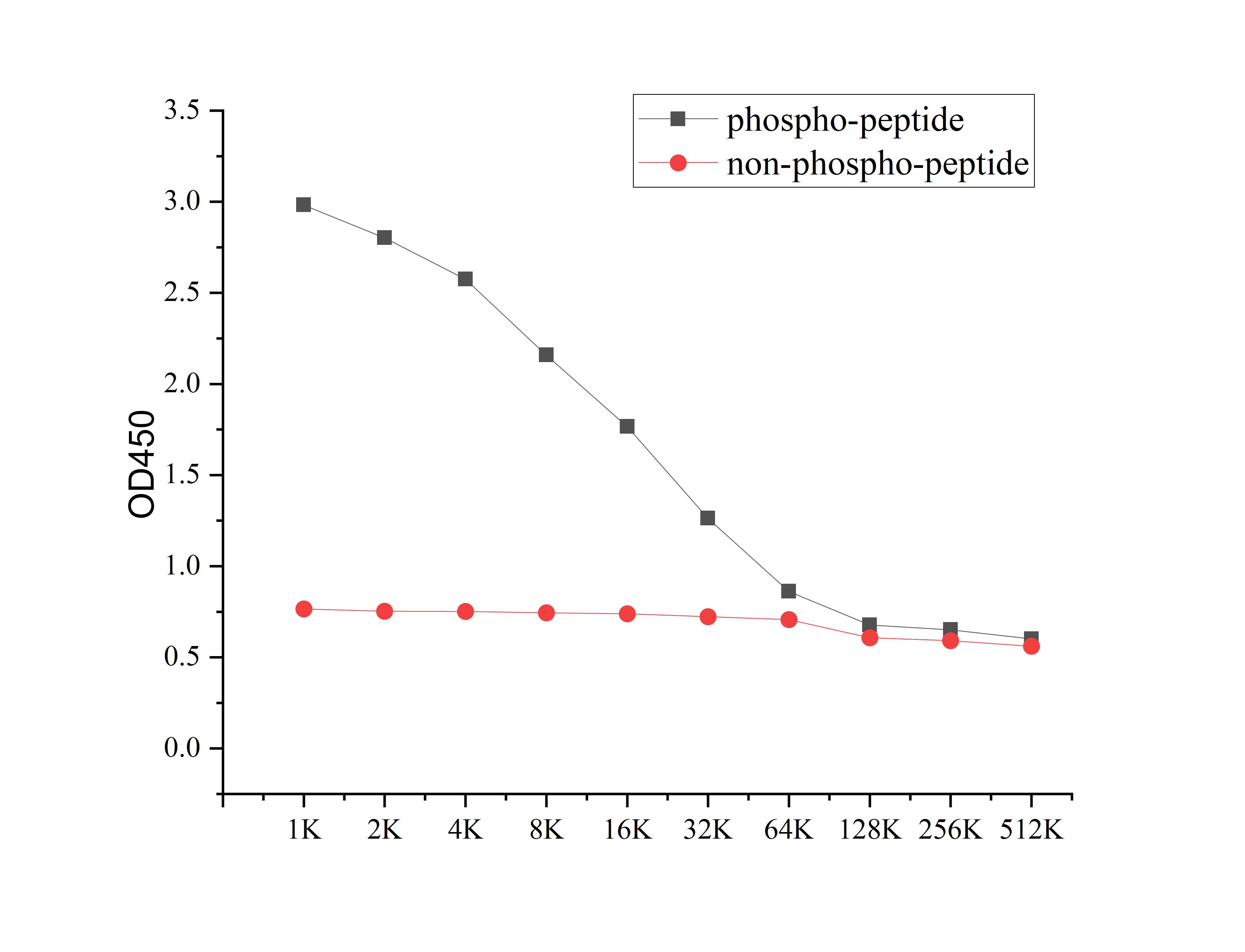
ELISA
Microtiter plate wells were coated with 100 ng/well coating antigen and incubated with serial dilutions of the antibody. Following HRP-conjugated secondary antibody and substrate development, OD630 was measured. The absorbance decreased progressively with increasing antibody dilution, yielding a sigmoidal titration curve that demonstrates the high antigen-binding activity of the polyclonal antibody.DB
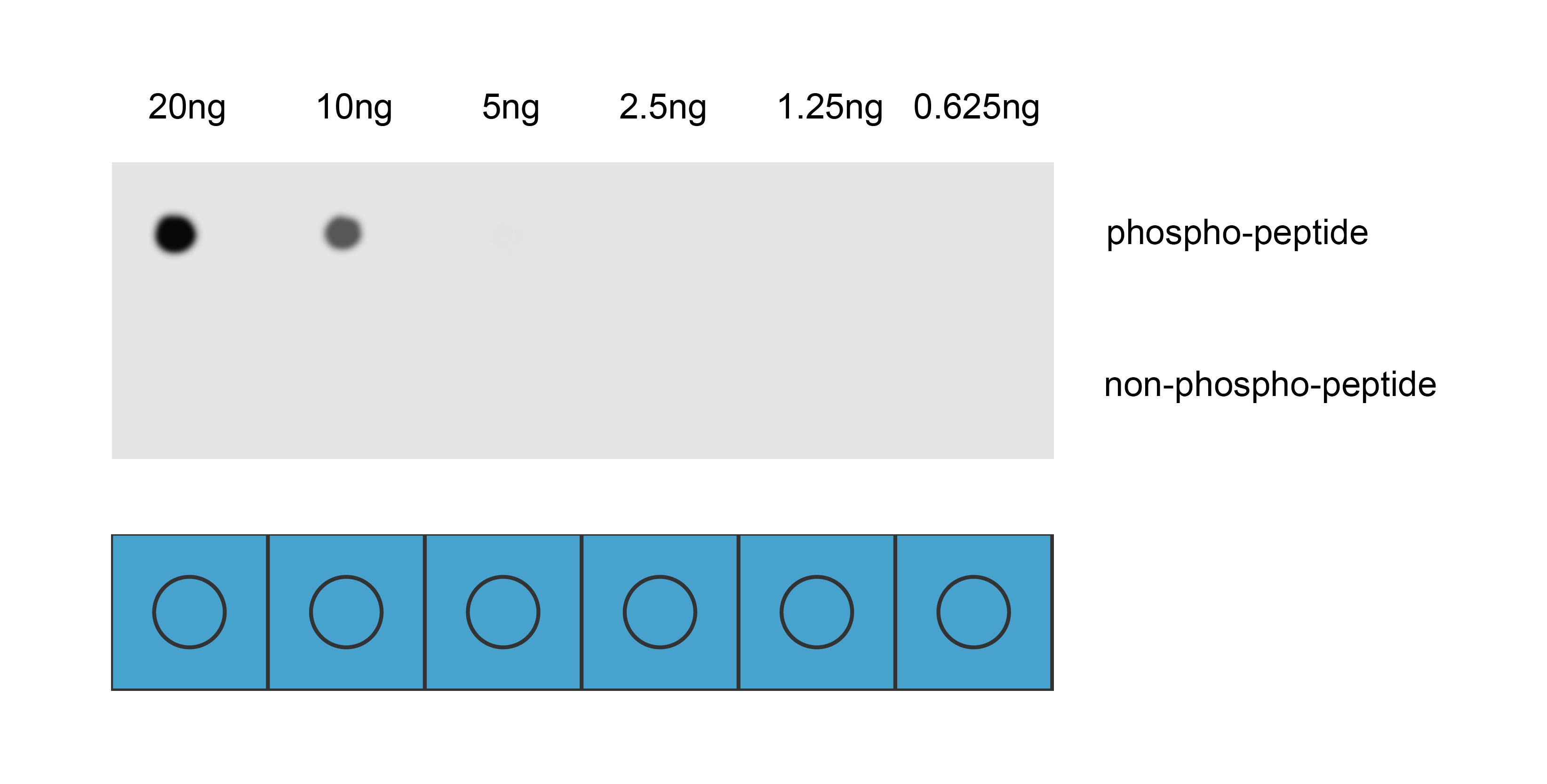
Dot blot analysis was performed using Anti-Phospho-LaminA/C(Ser392) antibody to detect two peptide samples: lane 1 contains the phospho-peptide , and lane 2 contains the non-phospho-peptide. Transfer the proteins onto a NC membrane (OM790001), and block it with TBST (OM750016) containing 5% BSA(OM297470) for 1 hour. Dilute the primary antibody with the antibody diluent (OM750012) at a ratio of 1:1000, and incubate it at room temperature for 2 hours. Wash the membrane 3 times with TBST (OM750016), 5 minutes each time. At room temperature, dilute the secondary antibody, Goat Anti-Rabbit IgG(H&L)-HRP (OM643487), at a ratio of 1:10000 and incubate for 1 hour. Wash the membrane 3 times with TBST (OM750016) again, 5 minutes each time. Use ECL (OM625701) for luminescence. Exposure time: 30 s.| Product Name | Anti-Phospho-LaminA/C(Ser392) antibody |
|---|---|
| Antibody Type | Primary Antibodies |
| Immunogen | Synthetic peptide conjugated with KLH |
| Modification | p-Ser392 |
| Clonality | Polyclonal |
|---|---|
| Isotype | IgG |
| Host Species | Rabbit |
| Tested Applications | DBELISA |
| DB:1:1000 |
|
| Concentration | 1mg/ml |
| Purification | Affinity purified |
| Gene Symbol | LMNA |
|---|---|
| Gene Synonyms | FPL IDC LFP CDDC EMD2 FPLD HGPS LDP1 LMN1 LMNC MADA PRO1 CDCD1 CMD1A FPLD2 LMNL1 CMT2B1 LGMD1B |
| Gene Full Name | lamin A/C |
| Gene Summary | The protein encoded by this gene is part of the nuclear lamina, a two-dimensional matrix of proteins located next to the inner nuclear membrane. The lamin family of proteins make up the matrix and are highly conserved in evolution. During mitosis, the lamina matrix is reversibly disassembled as the lamin proteins are phosphorylated. Lamin proteins are thought to be involved in nuclear stability, chromatin structure and gene expression. Vertebrate lamins consist of two types, A and B. Alternative splicing results in multiple transcript variants. Mutations in this gene lead to several diseases: Emery-Dreifuss muscular dystrophy, familial partial lipodystrophy, limb girdle muscular dystrophy, dilated cardiomyopathy, Charcot-Marie-Tooth disease, and Hutchinson-Gilford progeria syndrome. [provided by RefSeq, May 2022] |
| Alternative Names | 70 kDa lamin Cardiomyopathy dilated 1A (autosomal dominant) CDCD1 CDDC CMD1A CMT2B1 EMD2 FPL FPLD FPLD2 HGPS IDC Lamin A Lamin A/C Lamin A/C like 1 Lamin Lamin C Lamin-A/C LDP1 LFP LGMD1B Limb girdle muscular dystrophy 1B (autosomal dominant) LMN 1 LMN A LMN C LMN1 LMNA LMNA_HUMAN LMNC LMNL1 Prelamin A/C PRO1 Renal carcinoma antigen NY REN 32 Renal carcinoma antigen NY-REN-32 Renal carcinoma antigen NYREN32 |
| Molecular Weight(MW) | 74kDa |
| Cellular Localization | Nucleus. Nucleus envelope. Nucleus lamina. Nucleus>Nucleoplasm. |
| SwissProt ID | P02545 |
|---|
ELISA
Microtiter plate wells were coated with 100 ng/well coating antigen and incubated with serial dilutions of the antibody. Following HRP-conjugated secondary antibody and substrate development, OD630 was measured. The absorbance decreased progressively with increasing antibody dilution, yielding a sigmoidal titration curve that demonstrates the high antigen-binding activity of the polyclonal antibody.
DB
Dot blot analysis was performed using Anti-Phospho-LaminA/C(Ser392) antibody to detect two peptide samples: lane 1 contains the phospho-peptide , and lane 2 contains the non-phospho-peptide. Transfer the proteins onto a NC membrane (OM790001), and block it with TBST (OM750016) containing 5% BSA(OM297470) for 1 hour. Dilute the primary antibody with the antibody diluent (OM750012) at a ratio of 1:1000, and incubate it at room temperature for 2 hours. Wash the membrane 3 times with TBST (OM750016), 5 minutes each time. At room temperature, dilute the secondary antibody, Goat Anti-Rabbit IgG(H&L)-HRP (OM643487), at a ratio of 1:10000 and incubate for 1 hour. Wash the membrane 3 times with TBST (OM750016) again, 5 minutes each time. Use ECL (OM625701) for luminescence. Exposure time: 30 s.| Application Notes | DB:1:1000 |
|---|
| Form | Liquid |
|---|---|
| Storage Instructions | Shipped at 4°C. Store at +4°C short term (1-2 weeks). Store at -20°C long term. Avoid freeze / thaw cycle. |
| Storage Buffer | Purified antibody in PBS with 0.05% sodium azide,40% Glycerol. |


