ELISA
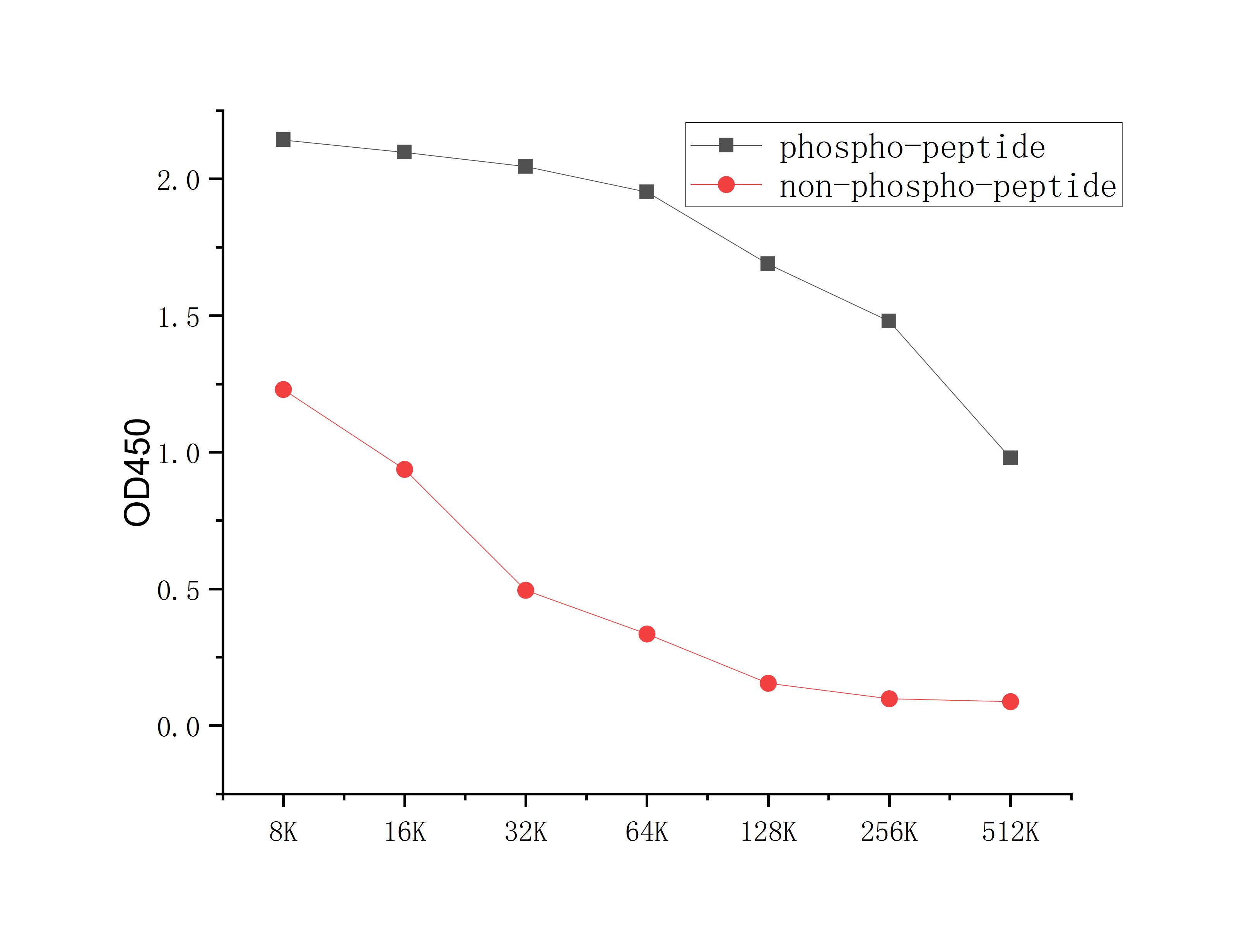
Coat the plate with 100ng immunizing antigen. The phospho-specific antibody,pbs only can specifically recognize phosphorylated peptide and enable quantitative detection over a wide dynamic range. Non-phosphorylated peptide shows no cross-reactivity, and the background signal remains low, indicating that the phospho-specific antibody,pbs only possesses high specificity and an excellent signal-to-noise ratio. Black line: phospho-peptide; Rad line: non-phospho peptideDB
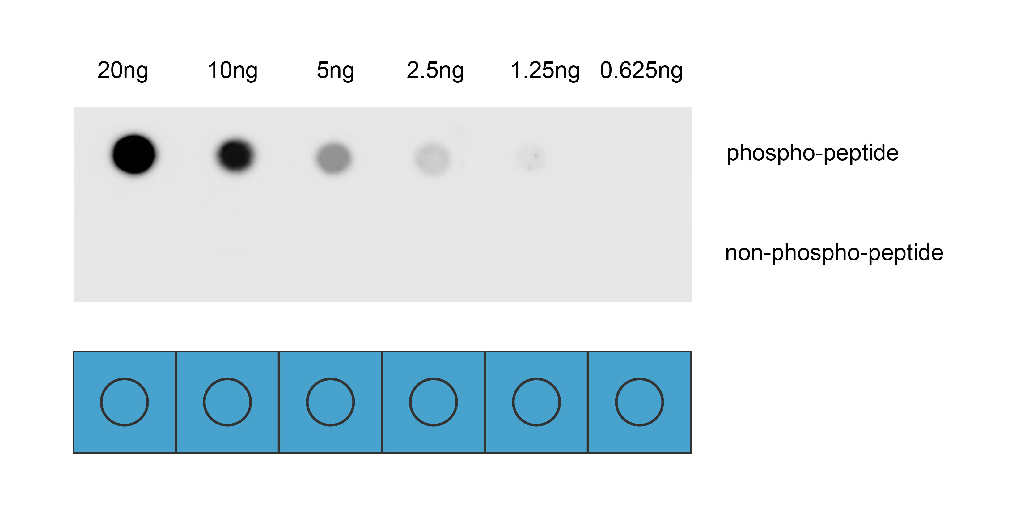
Dot blot analysis was performed using Anti-Phospho-c-Fos(Ser362) antibody to detect two peptide samples: lane 1 contains the phospho-peptide , and lane 2 contains the non-phospho-peptide. Transfer the proteins onto a NC membrane (OM790001), and block it with TBST (OM750016) containing 5% BSA(OM297470) for 1 hour. Dilute the primary antibody with the antibody diluent (OM750012) at a ratio of 1:1000, and incubate it at room temperature for 2 hours. Wash the membrane 3 times with TBST (OM750016), 5 minutes each time. At room temperature, dilute the secondary antibody, Goat Anti-Rabbit IgG(H&L)-HRP (OM643487), at a ratio of 1:10000 and incubate for 1 hour. Wash the membrane 3 times with TBST (OM750016) again, 5 minutes each time. Use ECL (OM625701) for luminescence. Exposure time: 30 s.WB
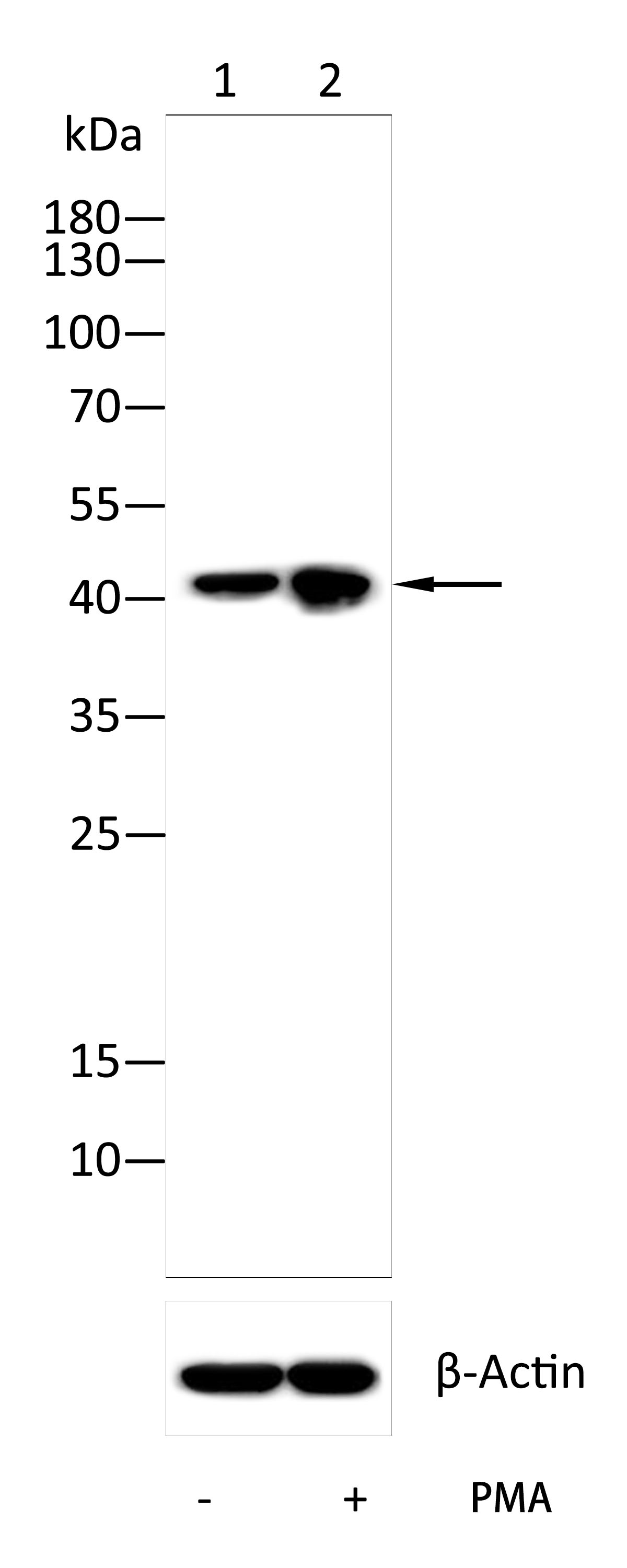
Western blot with Anti-Phospho-c-Fos(Ser362) antibody on lysates of NIH/3T3 cells (1) and NIH/3T3 cells serum-starved overnight and treated with 200 nM PMA (OM750071) for 30 min (2) .12% SDS-PAGE gel.Sample loading: 20μg /lane. Transfer the proteins onto a PVDF membrane (OM790003), and block it with TBST (OM750016) containing 5% BSA (OM297470) for one hour. Dilute the primary antibody with the antibody diluent (OM750012) at a ratio of 1:1000, and incubate it overnight at 4°C. Wash the membrane three times with TBST (OM750016), 5 minutes each time. At room temperature, dilute the secondary antibody, Goat Anti-Rabbit IgG(H&L)-HRP (OM643487), at a ratio of 1:10000 and incubate for one hour. Wash the membrane three times with TBST (OM750016) again, 5 minutes each time. Use ECL (OM625701) for luminescence.staining time: 120S.| Product Name | Anti-Phospho-c-Fos(Ser362) Antibody |
|---|---|
| Antibody Type | Primary Antibodies |
| Immunogen | Synthetic peptide conjugated with KLH. |
| Modification | p-Ser362 |
| Clonality | Polyclonal |
|---|---|
| Isotype | IgG |
| Host Species | Rabbit |
| Tested Applications | DBELISAWB |
| DB:1:1000 WB:1:1000 |
|
| Species Reactivity | Human |
| Concentration | 1mg/ml |
| Purification | Affinity purified |
| Gene Symbol | FOS |
|---|---|
| Gene Synonyms | p55 APhospho-1 C-FOS |
| Gene Full Name | Fos proto-oncogene, APhospho-1 transcription factor subunit |
| Gene Summary | The Fos gene family consists of 4 members: FOS, FOSB, FOSL1, and FOSL2. These genes encode leucine zipper proteins that can dimerize with proteins of the JUN family, thereby forming the transcription factor complex APhospho-1. As such, the FOS proteins have been implicated as regulators of cell proliferation, differentiation, and transformation. In some cases, expression of the FOS gene has also been associated with apoptotic cell death. |
| Alternative Names | Activator protein 1 antibody AP 1 antibody C FOS antibody Cellular oncogene c fos antibody Cellular oncogene fos antibody FBJ murine osteosarcoma viral (v fos) oncogene homolog (oncogene FOS) antibody FBJ murine osteosarcoma viral oncogene homolog antibody FBJ murine osteosarcoma viral v fos oncogene homolog antibody FBJ Osteosarcoma Virus antibody FOS antibody FOS protein antibody FOS_HUMAN antibody G0 G1 switch regulatory protein 7 antibody G0/G1 switch regulatory protein 7 antibody G0S7 antibody Oncogene FOS antibody p55 antibody proto oncogene c Fos antibody Proto oncogene protein c fos antibody Proto-oncogene c-Fos antibody v fos FBJ murine osteosarcoma viral oncogene homolog antibody |
| Molecular Weight(MW) | 41kDa(Predicted); 41kDa(Observed) |
| Cellular Localization | Nucleus, Endoplasmic reticulum, Cytoplasm, cytosol. |
| SwissProt ID | P01100 |
|---|
ELISA
Coat the plate with 100ng immunizing antigen. The phospho-specific antibody,pbs only can specifically recognize phosphorylated peptide and enable quantitative detection over a wide dynamic range. Non-phosphorylated peptide shows no cross-reactivity, and the background signal remains low, indicating that the phospho-specific antibody,pbs only possesses high specificity and an excellent signal-to-noise ratio. Black line: phospho-peptide; Rad line: non-phospho peptide
DB
Dot blot analysis was performed using Anti-Phospho-c-Fos(Ser362) antibody to detect two peptide samples: lane 1 contains the phospho-peptide , and lane 2 contains the non-phospho-peptide. Transfer the proteins onto a NC membrane (OM790001), and block it with TBST (OM750016) containing 5% BSA(OM297470) for 1 hour. Dilute the primary antibody with the antibody diluent (OM750012) at a ratio of 1:1000, and incubate it at room temperature for 2 hours. Wash the membrane 3 times with TBST (OM750016), 5 minutes each time. At room temperature, dilute the secondary antibody, Goat Anti-Rabbit IgG(H&L)-HRP (OM643487), at a ratio of 1:10000 and incubate for 1 hour. Wash the membrane 3 times with TBST (OM750016) again, 5 minutes each time. Use ECL (OM625701) for luminescence. Exposure time: 30 s.
WB
Western blot with Anti-Phospho-c-Fos(Ser362) antibody on lysates of NIH/3T3 cells (1) and NIH/3T3 cells serum-starved overnight and treated with 200 nM PMA (OM750071) for 30 min (2) .12% SDS-PAGE gel.Sample loading: 20μg /lane. Transfer the proteins onto a PVDF membrane (OM790003), and block it with TBST (OM750016) containing 5% BSA (OM297470) for one hour. Dilute the primary antibody with the antibody diluent (OM750012) at a ratio of 1:1000, and incubate it overnight at 4°C. Wash the membrane three times with TBST (OM750016), 5 minutes each time. At room temperature, dilute the secondary antibody, Goat Anti-Rabbit IgG(H&L)-HRP (OM643487), at a ratio of 1:10000 and incubate for one hour. Wash the membrane three times with TBST (OM750016) again, 5 minutes each time. Use ECL (OM625701) for luminescence.staining time: 120S.| Application Notes | DB:1:1000 WB:1:1000 |
|---|
| Form | Liquid |
|---|---|
| Storage Instructions | Shipped at 4°C. Store at +4°C short term (1-2 weeks). Store at -20°C long term. Avoid freeze / thaw cycle. |
| Storage Buffer | Purified antibody in PBS with 0.05% sodium azide,40% Glycerol. |


