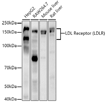
WB
Western blot analysis of various lysates using LDL Receptor (LDLR) Rabbit mAb at 1:1000 dilution. Secondary antibody: HRP-conjugated Goat anti-Rabbit IgG (H+L) at 1:10000 dilution. Lysates/proteins: 25μg per lane. Blocking buffer: 3% nonfat dry milk in TBST. Detection: ECL Basic Kit. Exposure time: 30s.IHC
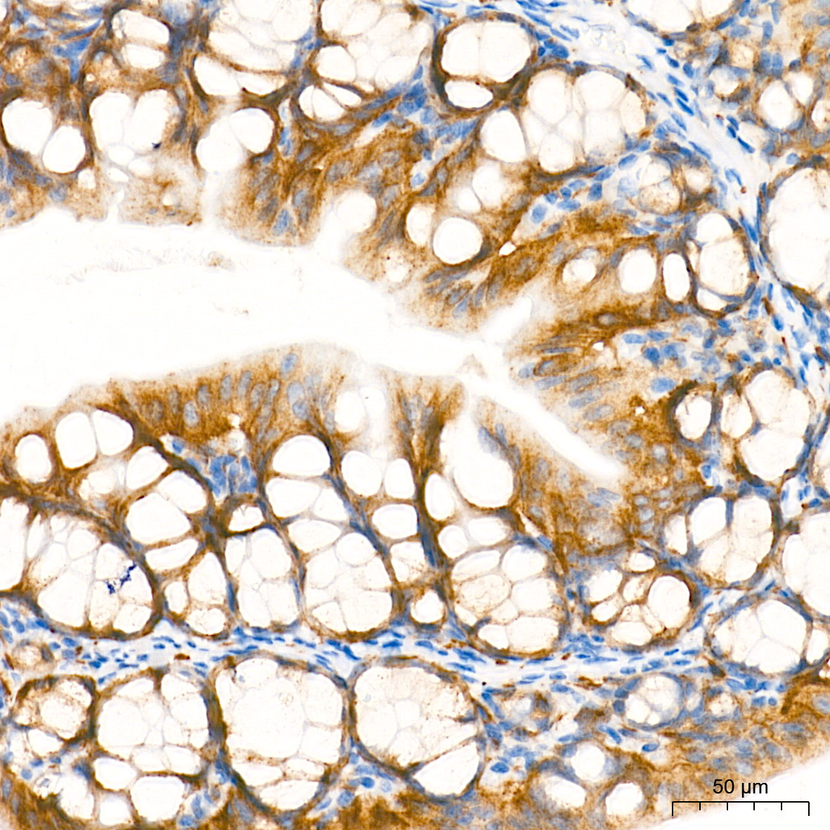
Immunohistochemistry analysis of paraffin embedded Mouse intestin tissue using LDL Receptor (LDLR) Rabbit mAb at a dilution of 1:600 (40x lens). High pressure antigen retrieval performed with 0.01M Tris EDTA Buffer (pH 9.0) prior to IHC staining.IHC
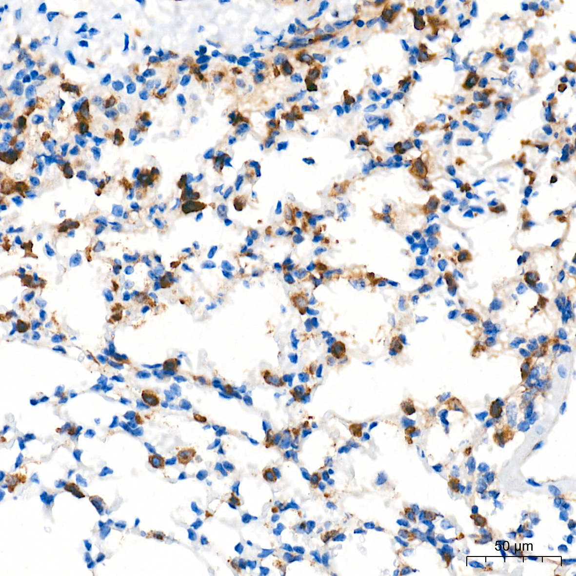
Immunohistochemistry analysis of paraffin embedded Mouse lung tissue using LDL Receptor (LDLR) Rabbit mAb at a dilution of 1:600 (40x lens). High pressure antigen retrieval performed with 0.01M Tris EDTA Buffer (pH 9.0) prior to IHC staining.ICC/IF
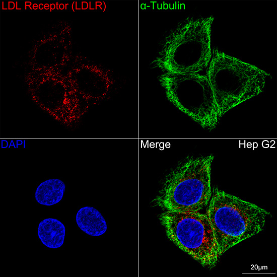
Confocal imaging of Hep G2 cells using LDL Receptor (LDLR) Rabbit mAb (dilution 1:200) followed by a further incubation with Cy3 Goat Anti-Rabbit IgG (H+L) (dilution 1:500) (Red). The cells were counterstained with α-Tubulin Mouse mAb (dilution 1:400) followed by incubation with Omnimabs® 488-conjugated Goat Anti-Mouse IgG (H+L) Ab (dilution 1:500) (Green). DAPI was used for nuclear staining (Blue). Objective: 100x.| Product Name | LDL Receptor (LDLR) Rabbit mAb |
|---|---|
| Antibody Type | Primary Antibodies |
| Immunogen | A synthetic peptide corresponding to a sequence within amino acids 761-860 of human LDL Receptor (LDLR) (NP_000518.1). |
| Clonality | monoclonal |
|---|---|
| Isotype | IgG |
| Host Species | Rabbit |
| Tested Applications | ICC/IFIHCWB |
| WB:1:1000-1:2000 IHC:1:100-1:500 ICC/IF:1:200-1:400 |
|
| Species Reactivity | HumanMouseRat |
| Concentration | 1mg/ml |
| Purification | Affinity purified |
| Gene Symbol | LDLR |
|---|---|
| Gene Synonyms | FH FHC FHCL1 LDLCQ2 |
| Gene Full Name | low density lipoprotein receptor |
| Gene Summary | The low density lipoprotein receptor (LDLR) gene family consists of cell surface proteins involved in receptor-mediated endocytosis of specific ligands. The encoded protein is normally bound at the cell membrane, where it binds low density lipoprotein/cholesterol and is taken into the cell. Lysosomes release the cholesterol, which is made available for repression of microsomal enzyme 3-hydroxy-3-methylglutaryl coenzyme A (HMG CoA) reductase, the rate-limiting step in cholesterol synthesis. At the same time, a reciprocal stimulation of cholesterol ester synthesis takes place. Mutations in this gene cause the autosomal dominant disorder, familial hypercholesterolemia. Alternate splicing results in multiple transcript variants.[provided by RefSeq, May 2022] |
| Molecular Weight(MW) | 95kDa(Observed MW 100-160kDa) |
| Cellular Localization | Cell membrane, Cell surface, Early endosome, Endomembrane system, Golgi apparatus, Late endosome, Lysosome, Membrane, Single-pass type I membrane protein, clathrin-coated pit. |
WB
Western blot analysis of various lysates using LDL Receptor (LDLR) Rabbit mAb at 1:1000 dilution. Secondary antibody: HRP-conjugated Goat anti-Rabbit IgG (H+L) at 1:10000 dilution. Lysates/proteins: 25μg per lane. Blocking buffer: 3% nonfat dry milk in TBST. Detection: ECL Basic Kit. Exposure time: 30s.
IHC
Immunohistochemistry analysis of paraffin embedded Mouse intestin tissue using LDL Receptor (LDLR) Rabbit mAb at a dilution of 1:600 (40x lens). High pressure antigen retrieval performed with 0.01M Tris EDTA Buffer (pH 9.0) prior to IHC staining.
IHC
Immunohistochemistry analysis of paraffin embedded Mouse lung tissue using LDL Receptor (LDLR) Rabbit mAb at a dilution of 1:600 (40x lens). High pressure antigen retrieval performed with 0.01M Tris EDTA Buffer (pH 9.0) prior to IHC staining.
ICC/IF
Confocal imaging of Hep G2 cells using LDL Receptor (LDLR) Rabbit mAb (dilution 1:200) followed by a further incubation with Cy3 Goat Anti-Rabbit IgG (H+L) (dilution 1:500) (Red). The cells were counterstained with α-Tubulin Mouse mAb (dilution 1:400) followed by incubation with Omnimabs® 488-conjugated Goat Anti-Mouse IgG (H+L) Ab (dilution 1:500) (Green). DAPI was used for nuclear staining (Blue). Objective: 100x.| Application Notes | WB:1:1000-1:2000 IHC:1:100-1:500 ICC/IF:1:200-1:400 |
|---|
| Form | Liquid |
|---|---|
| Storage Instructions | Store at -20℃. Avoid freeze / thaw cycles. |
| Storage Buffer | Buffer: PBS with 0.05% proclin300, 0.05% BSA, 50% glycerol, pH7.3. |


 Data sheet for OM644270