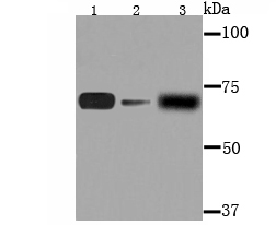
Fig1: Western blot analysis of AFP on different lysates using anti-AFP antibody at 1/1,000 dilution. Postive control: Lane 1: MCF-7 Lane 2: PC-12 Lane 3: Human liver tissue
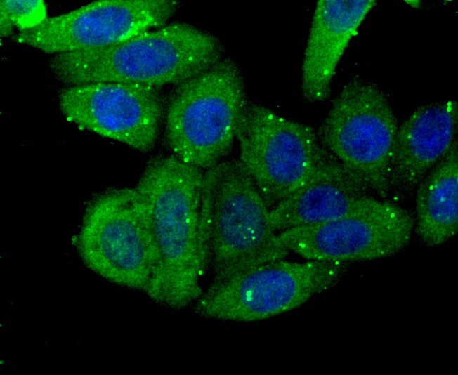
Fig2: ICC staining AFP in HepG2 cells (green). The nuclear counter stain is DAPI (blue). Cells were fixed in paraformaldehyde, permeabilised with 0.25% Triton X100/PBS.

Fig3: ICC staining AFP in MCF-7 cells (green). The nuclear counter stain is DAPI (blue). Cells were fixed in paraformaldehyde, permeabilised with 0.25% Triton X100/PBS.
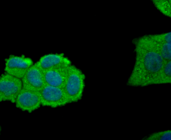
Fig4: ICC staining AFP in Hela cells (green). The nuclear counter stain is DAPI (blue). Cells were fixed in paraformaldehyde, permeabilised with 0.25% Triton X100/PBS.
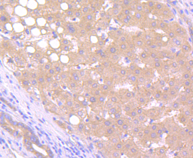
Fig5: Immunohistochemical analysis of paraffin-embedded human liver cancer tissue using anti-AFP antibody. Counter stained with hematoxylin.
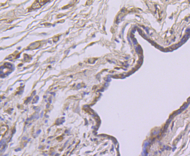
Fig6: Immunohistochemical analysis of paraffin-embedded human breast cancer tissue using anti-AFP antibody. Counter stained with hematoxylin.
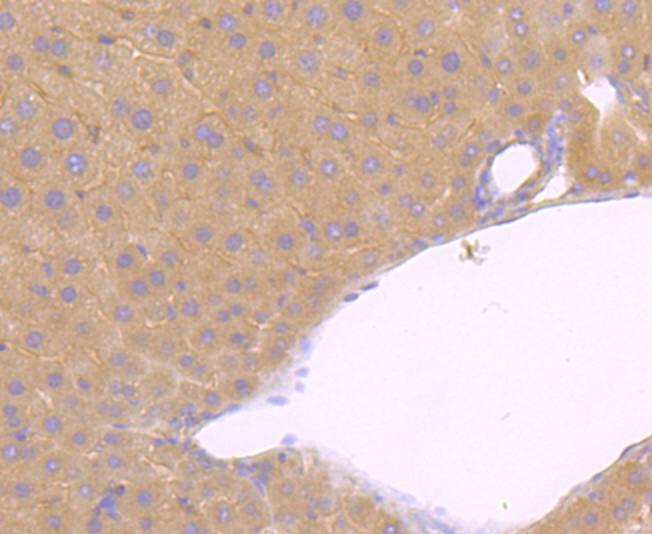
Fig7: Immunohistochemical analysis of paraffin-embedded mouse liver tissue using anti-AFP antibody. Counter stained with hematoxylin.
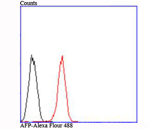
Fig8: Flow cytometric analysis of 239T cells with AFP antibody at 1/100 dilution (red) compared with an unlabelled control (cells without incubation with primary antibody; black).
Product Profile
| Product Name | AFP |
|---|---|
| Antibody Type | Primary Antibodies |
| Product description |
|
| Immunogen | Recombinant protein. |
Key Feature
| Clonality | Polyclonal |
|---|---|
| Isotype | IgG |
| Host Species | Rabbit |
| Tested Applications | |
WB:1:1,000-1:2,000 ICC:1:50-1:200 IHC:1:50-1:100 FC:1:50-1:100 |
|
| Species Reactivity | |
| Concentration | 1 mg/mL. |
Target Information
| Alternative Names | Afp antibody
AFPD antibody
Alpha fetoglobulin antibody
Alpha fetoprotein antibody
Alpha fetoprotein precursor antibody
Alpha-1-fetoprotein antibody
Alpha-fetoglobulin antibody
Alpha-fetoprotein antibody
alpha-fetoprotein Hereditary persistence of included antibody FETA antibody FETA_HUMAN antibody Hereditary persistence of alpha fetoprotein antibody HPAFP antibody |
|---|---|
| Cellular Localization | Secreted. |
Database Links
| SwissProt ID | P02771 P02772 P02773 |
|---|
Application
-

Application
Fig1: Western blot analysis of AFP on different lysates using anti-AFP antibody at 1/1,000 dilution. Postive control: Lane 1: MCF-7 Lane 2: PC-12 Lane 3: Human liver tissue
-

Application
Fig2: ICC staining AFP in HepG2 cells (green). The nuclear counter stain is DAPI (blue). Cells were fixed in paraformaldehyde, permeabilised with 0.25% Triton X100/PBS.
-

Application
Fig3: ICC staining AFP in MCF-7 cells (green). The nuclear counter stain is DAPI (blue). Cells were fixed in paraformaldehyde, permeabilised with 0.25% Triton X100/PBS.
-

Application
Fig4: ICC staining AFP in Hela cells (green). The nuclear counter stain is DAPI (blue). Cells were fixed in paraformaldehyde, permeabilised with 0.25% Triton X100/PBS.
-

Application
Fig5: Immunohistochemical analysis of paraffin-embedded human liver cancer tissue using anti-AFP antibody. Counter stained with hematoxylin.
-

Application
Fig6: Immunohistochemical analysis of paraffin-embedded human breast cancer tissue using anti-AFP antibody. Counter stained with hematoxylin.
-

Application
Fig7: Immunohistochemical analysis of paraffin-embedded mouse liver tissue using anti-AFP antibody. Counter stained with hematoxylin.
-

Application
Fig8: Flow cytometric analysis of 239T cells with AFP antibody at 1/100 dilution (red) compared with an unlabelled control (cells without incubation with primary antibody; black).
| Positive Control | MCF-7, PC-12 cell lysate, human liver tissue lysate, HepG2, Hela, human liver cancer tissue, human breast cancer tissue, mouse liver tissue, 293T. |
|---|---|
| Application Notes | WB:1:1,000-1:2,000 ICC:1:50-1:200 IHC:1:50-1:100 FC:1:50-1:100 |
Additional Information
| Form | Liquid |
|---|---|
| Storage Instructions | Store at +4℃ after thawing. Aliquot store at -20℃ or -80℃. Avoid repeated freeze / thaw cycles. |
| Storage Buffer | 1*TBS (pH7.4), 0.5%BSA, 50%Glycerol. Preservative: 0.05% Sodium Azide. |
- Related products
- Camel Growth Hormone (GH) ELISA Kit OM642709
- Camel Insulin-like growth factors 1 (IGF-1) ELISA Kit OM642708
- Human HIV-1 p24 core protein (HIV-1 p24) antibody ELISA Kit OM642706
- Antigen Repair 20X pH6.0 OM642703
- 647 labeled Tyramide OM642699
-
- ASSAY KITS
-
- SERUM
2013 © Omnimabs , All Rights Reserved.

