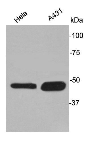
Fig1: Western blot analysis of Cytokeratin 17 on different lysates. Proteins were transferred to a PVDF membrane and blocked with 5% BSA in PBS for 1 hour at room temperature. The primary antibody was used at a 1:500 dilution in 5% BSA at room temperature for 2 hours. Goat Anti-Rabbit IgG - HRP Secondary Antibody (HA1001) at 1:5,000 dilution was used for 1 hour at room temperature.
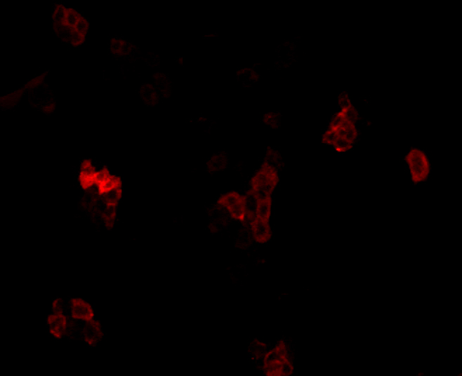
Fig2: ICC staining Cytokeratin 17 in A431 cells (red). Formalin fixed cells were permeabilized with 0.1% Triton X-100 in TBS for 10 minutes at room temperature and blocked with 1% Blocker BSA for 15 minutes at room temperature. Cells were probed with Cytokeratin 17 polyclonal antibody at a dilution of 1:100 for 1 hour at room temperature, washed with PBS. AlexaFluor®488 Goat anti-Rabbit IgG was used as the secondary antibody at 1/100 dilution.
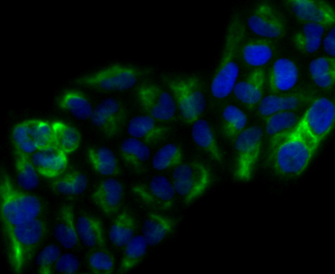
Fig3: ICC staining Cytokeratin 17 in Hela cells (green). Formalin fixed cells were permeabilized with 0.1% Triton X-100 in TBS for 10 minutes at room temperature and blocked with 1% Blocker BSA for 15 minutes at room temperature. Cells were probed with Cytokeratin 17 polyclonal antibody at a dilution of 1:100 for 1 hour at room temperature, washed with PBS. AlexaFluor®488 Goat anti-Rabbit IgG was used as the secondary antibody at 1/100 dilution. The nuclear counter stain is DAPI (blue).
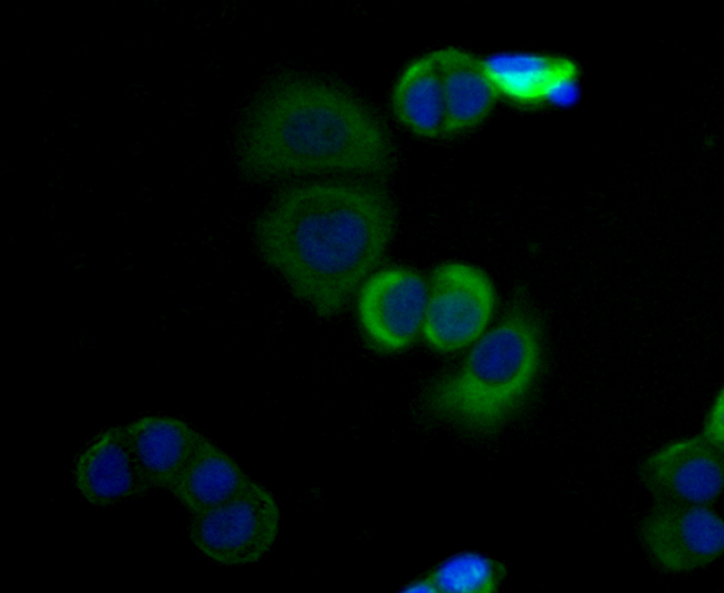
Fig4: ICC staining Cytokeratin 17 in SK-Br-3 cells (green). Formalin fixed cells were permeabilized with 0.1% Triton X-100 in TBS for 10 minutes at room temperature and blocked with 1% Blocker BSA for 15 minutes at room temperature. Cells were probed with Cytokeratin 17 polyclonal antibody at a dilution of 1:100 for 1 hour at room temperature, washed with PBS. AlexaFluor®488 Goat anti-Rabbit IgG was used as the secondary antibody at 1/100 dilution. The nuclear counter stain is DAPI (blue).
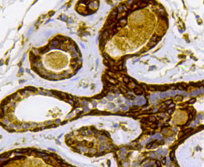
Fig5: Immunohistochemical analysis of paraffin-embedded human breast carcinoma tissue using anti-Cytokeratin 17 antibody. The section was pre-treated using heat mediated antigen retrieval with Tris-EDTA buffer (pH 8.0-8.4) for 20 minutes.The tissues were blocked in 5% BSA for 30 minutes at room temperature, washed with ddH2O and PBS, and then probed with 0407-4 at 1/100 dilution, for 30 minutes at room temperature and detected using an HRP conjugated compact polymer system. DAB was used as the chrogen. Counter stained with hematoxylin and mounted with DPX.
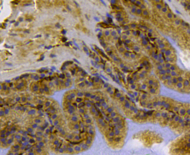
Fig6: Immunohistochemical analysis of paraffin-embedded mouse prostate tissue using anti-Cytokeratin 17 antibody. The section was pre-treated using heat mediated antigen retrieval with Tris-EDTA buffer (pH 8.0-8.4) for 20 minutes.The tissues were blocked in 5% BSA for 30 minutes at room temperature, washed with ddH2O and PBS, and then probed with 0407-4 at 1/100 dilution, for 30 minutes at room temperature and detected using an HRP conjugated compact polymer system. DAB was used as the chrogen. Counter stained with hematoxylin and mounted with DPX.
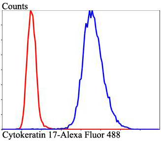
Fig7: Flow cytometric analysis of Cytokeratin 17 was done on Hela cells. The cells were fixed, permeabilized and stained with Cytokeratin 17 antibody at 1/100 dilution (red) compared with an unlabelled control (cells without incubation with primary antibody; blue). After incubation of the primary antibody on room temperature for an hour, the cells was stained with a Alexa Fluor 488-conjugated goat anti-rabbit IgG Secondary antibody at 1/500 dilution for at least 30 minutes.
Product Profile
| Product Name | Cytokeratin 17 |
|---|---|
| Antibody Type | Primary Antibodies |
| Product description |
|
| Immunogen | This antibody is produced by immunizing rabbits with a synthetic peptide (KLH-coupled) corresponding to near C-terminal residues of mouse CK-17. |
Key Feature
| Clonality | Polyclonal |
|---|---|
| Isotype | IgG |
| Host Species | Rabbit |
| Tested Applications | |
WB 1:2,000: ICC 1:50-1:200: IHC 1:50-1:200: FC 1:50-1:100: |
|
| Species Reactivity | |
| Concentration | 1 mg/mL. |
Target Information
| Alternative Names | 39.1 antibody
CK 17 antibody
CK-17 antibody
Cytokeratin-17 antibody
K17 antibody
K1C17_HUMAN antibody
Keratin 17 antibody
keratin 17 epitope S1 antibody
keratin 17 epitope S2 antibody
keratin 17 epitope S4 antibody
Keratin 17 type I antibody Keratin antibody Keratin type I cytoskeletal 17 antibody keratin type i cytoskeletal 17 [version 1] antibody Keratin-17 antibody KRT17 antibody PC antibody PC2 antibody PCHC1 antibody type I cytoskeletal 17 antibody |
|---|---|
| Molecular Weight(MW) | 48kDa |
| Cellular Localization | Cytoplasm |
Database Links
| SwissProt ID | Q04695 Q9QWL7 Q6IFU8 |
|---|
Application
-

Application
Fig1: Western blot analysis of Cytokeratin 17 on different lysates. Proteins were transferred to a PVDF membrane and blocked with 5% BSA in PBS for 1 hour at room temperature. The primary antibody was used at a 1:500 dilution in 5% BSA at room temperature for 2 hours. Goat Anti-Rabbit IgG - HRP Secondary Antibody (HA1001) at 1:5,000 dilution was used for 1 hour at room temperature.
-

Application
Fig2: ICC staining Cytokeratin 17 in A431 cells (red). Formalin fixed cells were permeabilized with 0.1% Triton X-100 in TBS for 10 minutes at room temperature and blocked with 1% Blocker BSA for 15 minutes at room temperature. Cells were probed with Cytokeratin 17 polyclonal antibody at a dilution of 1:100 for 1 hour at room temperature, washed with PBS. AlexaFluor®488 Goat anti-Rabbit IgG was used as the secondary antibody at 1/100 dilution.
-

Application
Fig3: ICC staining Cytokeratin 17 in Hela cells (green). Formalin fixed cells were permeabilized with 0.1% Triton X-100 in TBS for 10 minutes at room temperature and blocked with 1% Blocker BSA for 15 minutes at room temperature. Cells were probed with Cytokeratin 17 polyclonal antibody at a dilution of 1:100 for 1 hour at room temperature, washed with PBS. AlexaFluor®488 Goat anti-Rabbit IgG was used as the secondary antibody at 1/100 dilution. The nuclear counter stain is DAPI (blue).
-

Application
Fig4: ICC staining Cytokeratin 17 in SK-Br-3 cells (green). Formalin fixed cells were permeabilized with 0.1% Triton X-100 in TBS for 10 minutes at room temperature and blocked with 1% Blocker BSA for 15 minutes at room temperature. Cells were probed with Cytokeratin 17 polyclonal antibody at a dilution of 1:100 for 1 hour at room temperature, washed with PBS. AlexaFluor®488 Goat anti-Rabbit IgG was used as the secondary antibody at 1/100 dilution. The nuclear counter stain is DAPI (blue).
-

Application
Fig5: Immunohistochemical analysis of paraffin-embedded human breast carcinoma tissue using anti-Cytokeratin 17 antibody. The section was pre-treated using heat mediated antigen retrieval with Tris-EDTA buffer (pH 8.0-8.4) for 20 minutes.The tissues were blocked in 5% BSA for 30 minutes at room temperature, washed with ddH2O and PBS, and then probed with 0407-4 at 1/100 dilution, for 30 minutes at room temperature and detected using an HRP conjugated compact polymer system. DAB was used as the chrogen. Counter stained with hematoxylin and mounted with DPX.
-

Application
Fig6: Immunohistochemical analysis of paraffin-embedded mouse prostate tissue using anti-Cytokeratin 17 antibody. The section was pre-treated using heat mediated antigen retrieval with Tris-EDTA buffer (pH 8.0-8.4) for 20 minutes.The tissues were blocked in 5% BSA for 30 minutes at room temperature, washed with ddH2O and PBS, and then probed with 0407-4 at 1/100 dilution, for 30 minutes at room temperature and detected using an HRP conjugated compact polymer system. DAB was used as the chrogen. Counter stained with hematoxylin and mounted with DPX.
-

Application
Fig7: Flow cytometric analysis of Cytokeratin 17 was done on Hela cells. The cells were fixed, permeabilized and stained with Cytokeratin 17 antibody at 1/100 dilution (red) compared with an unlabelled control (cells without incubation with primary antibody; blue). After incubation of the primary antibody on room temperature for an hour, the cells was stained with a Alexa Fluor 488-conjugated goat anti-rabbit IgG Secondary antibody at 1/500 dilution for at least 30 minutes.
| Positive Control | Hela, A431, SK-Br-3, human breast carcinoma tissue, mouse prostate tissue. |
|---|---|
| Application Notes | WB 1:2,000: ICC 1:50-1:200: IHC 1:50-1:200: FC 1:50-1:100: |
Additional Information
| Form | Liquid |
|---|---|
| Storage Instructions | Store at +4℃ after thawing. Aliquot store at -20℃. Avoid repeated freeze / thaw cycles. |
| Storage Buffer | 1*TBS (pH7.4), 1%BSA, 50%Glycerol. Preservative: 0.05% Sodium Azide. |
- Related products
- Human ALANINE ELISA Kit OM642712
- Human β-ALANINE ELISA Kit OM642711
- Camel Growth Hormone (GH) ELISA Kit OM642709
- Camel Insulin-like growth factors 1 (IGF-1) ELISA Kit OM642708
- Human HIV-1 p24 core protein (HIV-1 p24) antibody ELISA Kit OM642706
-
- ASSAY KITS
-
- SERUM
2013 © Omnimabs , All Rights Reserved.

