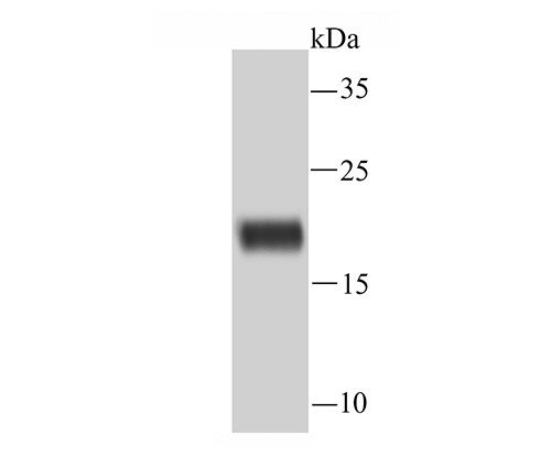
Fig1: Western blot analysis of Ferritin on rat spleen tissue lysate using anti-Ferritin antibody at 1/1,000 dilution.
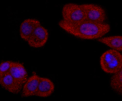
Fig2: ICC staining Ferritin in MCF-7 cells (red). The nuclear counter stain is DAPI (blue). Cells were fixed in paraformaldehyde, permeabilised with 0.25% Triton X100/PBS.
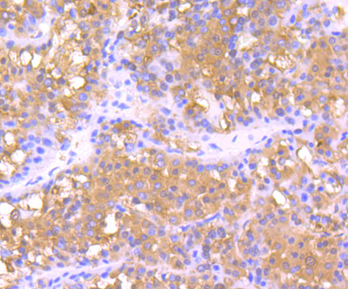
Fig3: Immunohistochemical analysis of paraffin-embedded human liver cancer tissue using anti-Ferritin antibody. Counter stained with hematoxylin.
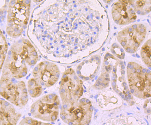
Fig4: Immunohistochemical analysis of paraffin-embedded human kidney tissue using anti-Ferritin antibody. Counter stained with hematoxylin.
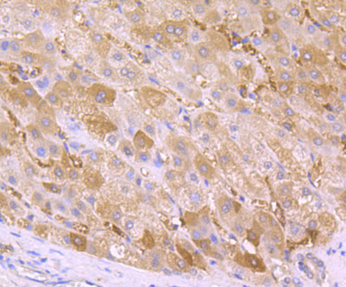
Fig5: Immunohistochemical analysis of paraffin-embedded human liver tissue using anti-Ferritin antibody. Counter stained with hematoxylin.
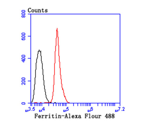
Fig6: Flow cytometric analysis of HepG2 cells with Ferritin antibody at 1/100 dilution (red) compared with an unlabelled control (cells without incubation with primary antibody; black).
Product Profile
| Product Name | Ferritin |
|---|---|
| Antibody Type | Primary Antibodies |
| Product description |
|
| Immunogen | Recombinant protein |
Key Feature
| Clonality | Polyclonal |
|---|---|
| Isotype | IgG |
| Host Species | Rabbit |
| Tested Applications | |
WB:1:500~1:2000 ICC:1:200~1:500 IHC:1:50~1:200 FC:1:200~1:500 Notes:Optimal dilutions/concentrations should be determined by the researcher. |
|
| Species Reactivity | |
| Concentration | 1 mg/mL. |
Target Information
| Alternative Names | Cell proliferation-inducing gene 15 protein antibody
Ferritin H subunit antibody
Ferritin heavy chain antibody
Ferritin heavy polypeptide 1 antibody
Ferritin L subunit antibody
Ferritin light polypeptide antibody
Ferritin heavy polypeptide antibody FRIH_HUMAN antibody FTH antibody FTH1 antibody FTL antibody |
|---|---|
| Molecular Weight(MW) | 21 kDa |
| Cellular Localization | Cytoplasm. |
Database Links
| SwissProt ID | P02792 P02794 P02793 P19132 |
|---|
Application
-

Application
Fig1: Western blot analysis of Ferritin on rat spleen tissue lysate using anti-Ferritin antibody at 1/1,000 dilution.
-

Application
Fig2: ICC staining Ferritin in MCF-7 cells (red). The nuclear counter stain is DAPI (blue). Cells were fixed in paraformaldehyde, permeabilised with 0.25% Triton X100/PBS.
-

Application
Fig3: Immunohistochemical analysis of paraffin-embedded human liver cancer tissue using anti-Ferritin antibody. Counter stained with hematoxylin.
-

Application
Fig4: Immunohistochemical analysis of paraffin-embedded human kidney tissue using anti-Ferritin antibody. Counter stained with hematoxylin.
-

Application
Fig5: Immunohistochemical analysis of paraffin-embedded human liver tissue using anti-Ferritin antibody. Counter stained with hematoxylin.
-

Application
Fig6: Flow cytometric analysis of HepG2 cells with Ferritin antibody at 1/100 dilution (red) compared with an unlabelled control (cells without incubation with primary antibody; black).
| Positive Control | Rat spleen tissue lysate, human liver tissue, human liver cancer tissue, human kidney tissue, MCF-7, HepG2. |
|---|---|
| Application Notes | WB:1:500~1:2000 ICC:1:200~1:500 IHC:1:50~1:200 FC:1:200~1:500 Notes:Optimal dilutions/concentrations should be determined by the researcher. |
Additional Information
| Form | Liquid |
|---|---|
| Storage Instructions | Store at +4℃ after thawing. Aliquot store at -20℃ or -80℃. Avoid repeated freeze / thaw cycles. |
| Storage Buffer | 1*TBS (pH7.4), 0.5%BSA, 50%Glycerol. Preservative: 0.05% Sodium Azide. |
- Related products
- Human ALANINE ELISA Kit OM642712
- Human β-ALANINE ELISA Kit OM642711
- Camel Growth Hormone (GH) ELISA Kit OM642709
- Camel Insulin-like growth factors 1 (IGF-1) ELISA Kit OM642708
- Human HIV-1 p24 core protein (HIV-1 p24) antibody ELISA Kit OM642706
-
- ASSAY KITS
-
- SERUM
2013 © Omnimabs , All Rights Reserved.

