
Fig1: Western blot analysis of GRP78 on different cell lysates using anti-GRP78 antibody at 1/2000 dilution.
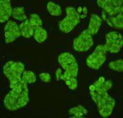
Fig2: ICC staining GRP78 in Hela cells (green). Cells were fixed in paraformaldehyde, permeabilised with 0.25% Triton X100/PBS.
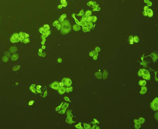
Fig3: ICC staining GRP78 in SK-BR-3 cells (green). Cells were fixed in paraformaldehyde, permeabilised with 0.25% Triton X100/PBS.

Fig4: ICC staining GRP78 in HepG2 cells (green). Cells were fixed in paraformaldehyde, permeabilised with 0.25% Triton X100/PBS.
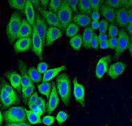
Fig5: IF staining GRP78 in Hela cells (green). Cells were fixed in paraformaldehyde, permeabilised with 0.25% Triton X100/PBS and counterstained with DAPI in order to highlight the nucleus (blue).
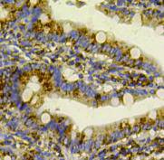
Fig6: Immunohistochemical analysis of paraffin-embedded rat small intestine tissue using anti-GRP78 antibody. Counter stained with hematoxylin.
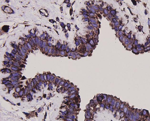
Fig7: Immunohistochemical analysis of paraffin-embedded human breast carcinoma tissue using anti-GRP78 antibody. Counter stained with hematoxylin.
Product Profile
| Product Name | GRP78 |
|---|---|
| Antibody Type | Primary Antibodies |
| Product description |
|
| Immunogen | peptide |
Key Feature
| Clonality | Polyclonal |
|---|---|
| Isotype | IgG |
| Host Species | Rabbit |
| Tested Applications | |
WB:1:2,000 ICC:1:200 IHC:1:200 FC:1:100-1:200 |
|
| Species Reactivity | |
| Concentration | 1 mg/mL. |
Target Information
| Alternative Names | 78 kDa glucose regulated protein antibody
78 kDa glucose-regulated protein antibody
AL022860 antibody
AU019543 antibody
BIP antibody
D2Wsu141e antibody
D2Wsu17e antibody
Endoplasmic reticulum lumenal Ca(2+)-binding protein grp78 antibody
Endoplasmic reticulum lumenal Ca2+ binding protein grp78 antibody
Epididymis secretory sperm binding protein Li 89n antibody
FLJ26106 antibody
Glucose Regulated Protein 78kDa antibody
GRP 78 antibody
GRP-78 antibody
GRP78 antibody
GRP78_HUMAN antibody
Heat shock 70 kDa protein 5 antibody
Heat Shock 70kDa Protein 5 antibody
Heat shock protein family A (Hsp70) member 5 antibody
HEL S 89n antibody
Hsce70 antibody
HSPA 5 antibody
HSPA5 antibody
Immunoglobulin Heavy Chain Binding Protein antibody
Immunoglobulin heavy chain-binding protein antibody
mBiP antibody
MIF2 antibody
Sez7 antibody
|
|---|---|
| Molecular Weight(MW) | 72 kDa |
| Cellular Localization | Cytoplasm, endoplasmic reticulum lumen |
Database Links
| SwissProt ID | P11021 |
|---|
Application
-

Application
Fig1: Western blot analysis of GRP78 on different cell lysates using anti-GRP78 antibody at 1/2000 dilution.
-

Application
Fig2: ICC staining GRP78 in Hela cells (green). Cells were fixed in paraformaldehyde, permeabilised with 0.25% Triton X100/PBS.
-

Application
Fig3: ICC staining GRP78 in SK-BR-3 cells (green). Cells were fixed in paraformaldehyde, permeabilised with 0.25% Triton X100/PBS.
-

Application
Fig4: ICC staining GRP78 in HepG2 cells (green). Cells were fixed in paraformaldehyde, permeabilised with 0.25% Triton X100/PBS.
-

Application
Fig5: IF staining GRP78 in Hela cells (green). Cells were fixed in paraformaldehyde, permeabilised with 0.25% Triton X100/PBS and counterstained with DAPI in order to highlight the nucleus (blue).
-

Application
Fig6: Immunohistochemical analysis of paraffin-embedded rat small intestine tissue using anti-GRP78 antibody. Counter stained with hematoxylin.
-

Application
Fig7: Immunohistochemical analysis of paraffin-embedded human breast carcinoma tissue using anti-GRP78 antibody. Counter stained with hematoxylin.
| Positive Control | Hela, PC12, HepG2, SK-BR-3, human liver tissue, rat epencephalon tissue, mouse liver tissue, rat small intestine tissue, mouse small intestine, human colon cancer tissue, human breast carcinoma tissue. |
|---|---|
| Application Notes | WB:1:2,000 ICC:1:200 IHC:1:200 FC:1:100-1:200 |
Additional Information
| Form | Liquid |
|---|---|
| Storage Instructions | Store at +4℃ after thawing. Aliquot store at -20℃ or -80℃. Avoid repeated freeze / thaw cycles. |
| Storage Buffer | 1*TBS (pH7.4), 1%BSA, 40%Glycerol. Preservative: 0.05% Sodium Azide. |
- Related products
- Rabbit Anti-Staphyloccocus aureus antibody OM642717
- Goat Anti-Rat IgG H&L (Alexa Fluor® 488) preadsorbed OM642716
- Goat Anti-Rabbit IgG H&L (Alexa Fluor® 488) OM642715
- Human ALANINE ELISA Kit OM642712
- Human β-ALANINE ELISA Kit OM642711
-
- ASSAY KITS
-
- SERUM
2013 © Omnimabs , All Rights Reserved.

