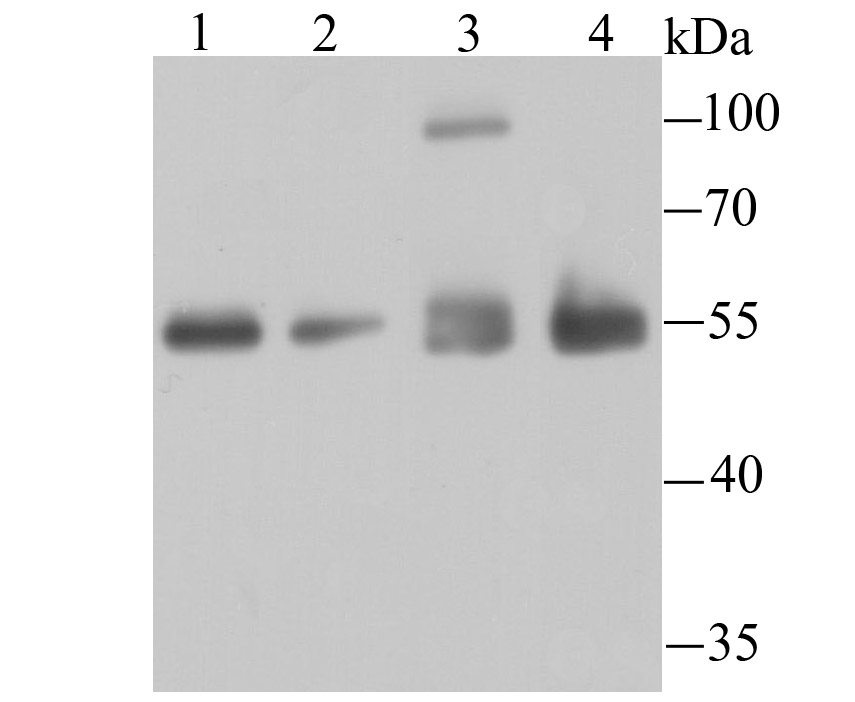
Fig1: Western blot analysis of HDAC2 on different cell lysates using anti-HDAC2 antibody at 1/1,000 dilution. Positive control: Lane1: SH-SY5Y Lane2: 293T Lane3: Hela Lane4: PC-12
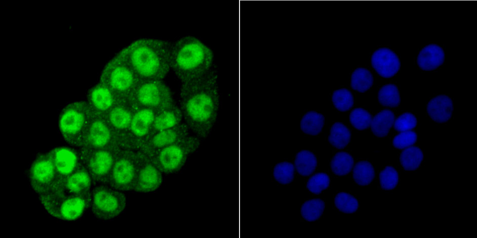
Fig2: ICC staining HDAC2 in LOVO cells (green). The nuclear counter stain is DAPI (blue). Cells were fixed in paraformaldehyde, permeabilised with 0.25% Triton X100/PBS.
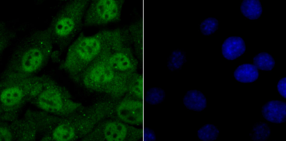
Fig3: ICC staining HDAC2 in NIH-3T3 cells (green). The nuclear counter stain is DAPI (blue). Cells were fixed in paraformaldehyde, permeabilised with 0.25% Triton X100/PBS.
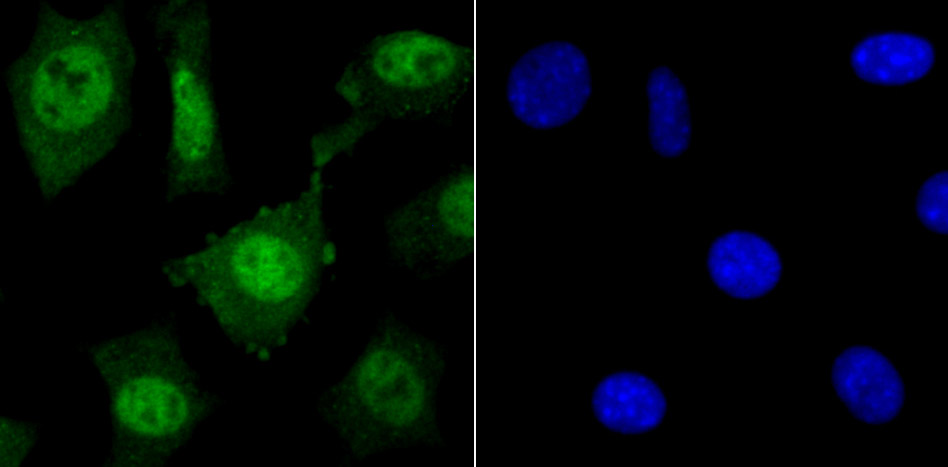
Fig4: ICC staining HDAC2 in SH-SY5Y cells (green). The nuclear counter stain is DAPI (blue). Cells were fixed in paraformaldehyde, permeabilised with 0.25% Triton X100/PBS.
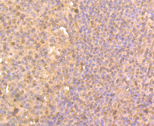
Fig5: Immunohistochemical analysis of paraffin-embedded human tonsil tissue using anti-HDAC2 antibody. Counter stained with hematoxylin.
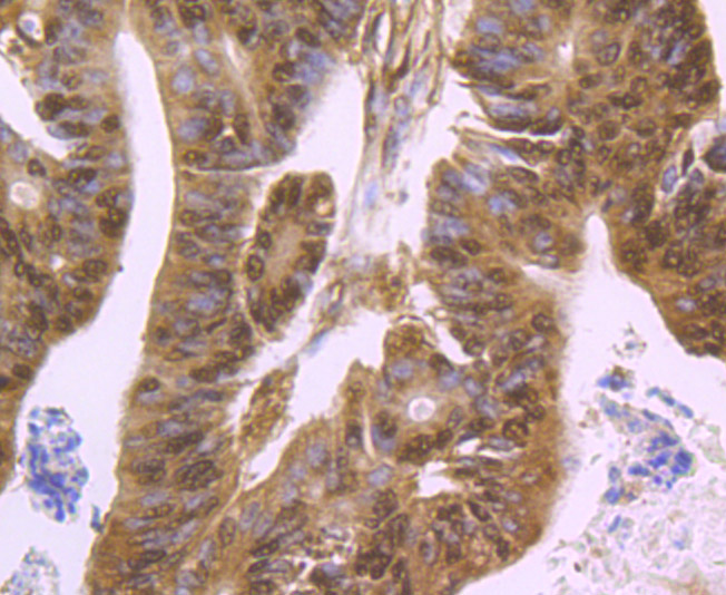
Fig6: Immunohistochemical analysis of paraffin-embedded human colon cancer tissue using anti-HDAC2 antibody. Counter stained with hematoxylin.
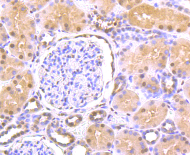
Fig7: Immunohistochemical analysis of paraffin-embedded human kidney tissue using anti-HDAC2 antibody. Counter stained with hematoxylin.
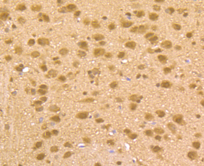
Fig8: Immunohistochemical analysis of paraffin-embedded mouse brain tissue using anti-HDAC2 antibody. Counter stained with hematoxylin.
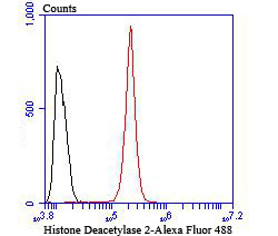
Fig9: Flow cytometric analysis of SH-SY5Y cells with HDAC2 antibody at 1/100 dilution (red) compared with an unlabelled control (cells without incubation with primary antibody; black).
Product Profile
| Product Name | Histone Deacetylase 2 |
|---|---|
| Antibody Type | Primary Antibodies |
| Product description |
|
| Immunogen | Recombinant protein. |
Key Feature
| Clonality | Polyclonal |
|---|---|
| Isotype | IgG |
| Host Species | Rabbit |
| Tested Applications | |
WB:1:500-1:1,000 ICC:1:500-1:2,000 IHC:1:100-1:200 FC:1:50-1:100 |
|
| Species Reactivity | |
| Concentration | 1 mg/mL. |
Target Information
| Alternative Names | D10Wsu179e antibody
HD 2 antibody
HD2 antibody
HDAC 2 antibody
Hdac2 antibody
HDAC2_HUMAN antibody
Histone deacetylase 2 (HD2) antibody
Histone deacetylase 2 antibody
OTTHUMP00000017046 antibody
OTTHUMP00000227077 antibody
OTTHUMP00000227078 antibody
RPD3 antibody
transcriptional regulator homolog RPD3 antibody
YAF1 antibody
YY1 associated factor 1 antibody
YY1 transcription factor binding protein antibody
Yy1bp antibody
|
|---|---|
| Molecular Weight(MW) | 55 kDa |
| Cellular Localization | Nucleus. Cytoplasm. |
Database Links
| SwissProt ID | Q92769 P70288 B1WBY8 |
|---|
Application
-

Application
Fig1: Western blot analysis of HDAC2 on different cell lysates using anti-HDAC2 antibody at 1/1,000 dilution. Positive control: Lane1: SH-SY5Y Lane2: 293T Lane3: Hela Lane4: PC-12
-

Application
Fig2: ICC staining HDAC2 in LOVO cells (green). The nuclear counter stain is DAPI (blue). Cells were fixed in paraformaldehyde, permeabilised with 0.25% Triton X100/PBS.
-

Application
Fig3: ICC staining HDAC2 in NIH-3T3 cells (green). The nuclear counter stain is DAPI (blue). Cells were fixed in paraformaldehyde, permeabilised with 0.25% Triton X100/PBS.
-

Application
Fig4: ICC staining HDAC2 in SH-SY5Y cells (green). The nuclear counter stain is DAPI (blue). Cells were fixed in paraformaldehyde, permeabilised with 0.25% Triton X100/PBS.
-

Application
Fig5: Immunohistochemical analysis of paraffin-embedded human tonsil tissue using anti-HDAC2 antibody. Counter stained with hematoxylin.
-

Application
Fig6: Immunohistochemical analysis of paraffin-embedded human colon cancer tissue using anti-HDAC2 antibody. Counter stained with hematoxylin.
-

Application
Fig7: Immunohistochemical analysis of paraffin-embedded human kidney tissue using anti-HDAC2 antibody. Counter stained with hematoxylin.
-

Application
Fig8: Immunohistochemical analysis of paraffin-embedded mouse brain tissue using anti-HDAC2 antibody. Counter stained with hematoxylin.
-

Application
Fig9: Flow cytometric analysis of SH-SY5Y cells with HDAC2 antibody at 1/100 dilution (red) compared with an unlabelled control (cells without incubation with primary antibody; black).
| Positive Control | SH-SY5Y, 293T, Hela, PC-12, LOVO, NIH-3T3, human tonsil tissue, human colon cancer tissue, human kidney tissue, mouse brain tissue. |
|---|---|
| Application Notes | WB:1:500-1:1,000 ICC:1:500-1:2,000 IHC:1:100-1:200 FC:1:50-1:100 |
Additional Information
| Form | Liquid |
|---|---|
| Storage Instructions | Store at +4℃ after thawing. Aliquot store at -20℃ or -80℃. Avoid repeated freeze / thaw cycles. |
| Storage Buffer | 1*TBS (pH7.4), 0.5%BSA, 50%Glycerol. Preservative: 0.05% Sodium Azide. |
- Related products
- Camel Growth Hormone (GH) ELISA Kit OM642709
- Camel Insulin-like growth factors 1 (IGF-1) ELISA Kit OM642708
- Human HIV-1 p24 core protein (HIV-1 p24) antibody ELISA Kit OM642706
- Antigen Repair 20X pH6.0 OM642703
- 647 labeled Tyramide OM642699
-
- ASSAY KITS
-
- SERUM
2013 © Omnimabs , All Rights Reserved.

