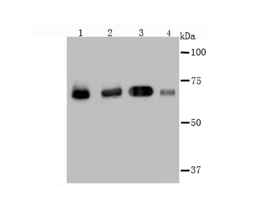
Fig1: Western blot analysis of Hsc70 on different cell lysate using anti-Hsc70 antibody at 1/2,000 dilution. Positive control:Lane1: HelaLane2: A431Lane3: NIH-3T3Lane4: PC-12
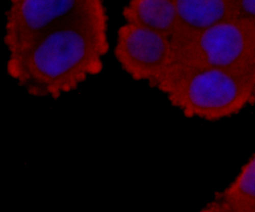
Fig2: ICC staining Hsc70 in Hela cells (red). The nuclear counter stain is DAPI (blue). Cells were fixed in paraformaldehyde, permeabilised with 0.25% Triton X100/PBS.
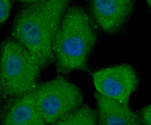
Fig3: ICC staining Hsc70 in A549 cells (green). The nuclear counter stain is DAPI (blue). Cells were fixed in paraformaldehyde, permeabilised with 0.25% Triton X100/PBS.
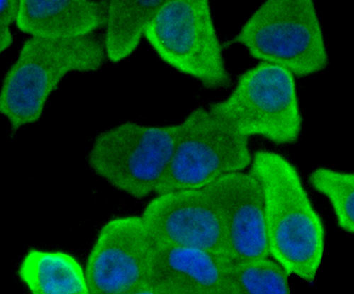
Fig4: ICC staining Hsc70 in SK-Br-3 cells (green). The nuclear counter stain is DAPI (blue). Cells were fixed in paraformaldehyde, permeabilised with 0.25% Triton X100/PBS.
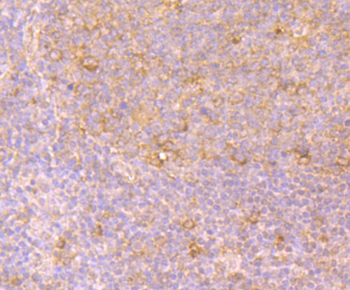
Fig5: Immunohistochemical analysis of paraffin-embedded human tonsil tissue using anti-Hsc70 antibody. Counter stained with hematoxylin.
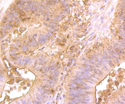
Fig6: Immunohistochemical analysis of paraffin-embedded human colon cancer tissue using anti-Hsc70 antibody. Counter stained with hematoxylin.
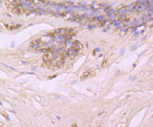
Fig7: Immunohistochemical analysis of paraffin-embedded human breast tissue using anti-Hsc70 antibody. Counter stained with hematoxylin.
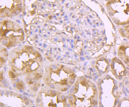
Fig8: Immunohistochemical analysis of paraffin-embedded human kidney tissue using anti-Hsc70 antibody. Counter stained with hematoxylin.
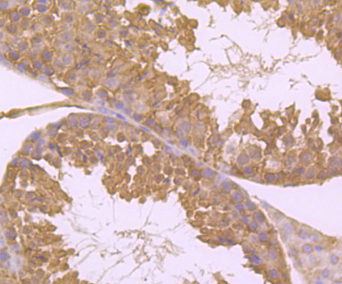
Fig9: Immunohistochemical analysis of paraffin-embedded mouse testis tissue using anti-Hsc70 antibody. Counter stained with hematoxylin.
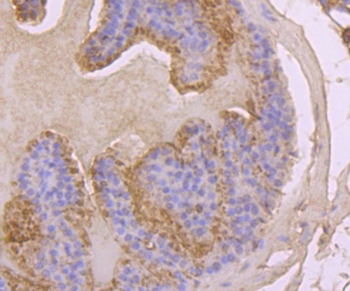
Fig10: Immunohistochemical analysis of paraffin-embedded mouse prostate tissue using anti-Hsc70 antibody. Counter stained with hematoxylin.
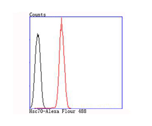
Fig11: Flow cytometric analysis of Jurkat cells with Hsc70 antibody at 1/100 dilution (red) compared with an unlabelled control (cells without incubation with primary antibody; black).
Product Profile
| Product Name | Hsc70 |
|---|---|
| Antibody Type | Primary Antibodies |
| Product description |
|
| Immunogen | Recombinant protein. |
Key Feature
| Clonality | Polyclonal |
|---|---|
| Isotype | IgG |
| Host Species | Rabbit |
| Tested Applications | |
WB:1:500~1:2000 ICC:1:200~1:500 IHC:1:50~1:200 FC:1:200~1:500 Notes:Optimal dilutions/concentrations should be determined by the researcher. |
|
| Species Reactivity | |
| Concentration | 1 mg/mL. |
Target Information
| Alternative Names | 2410008N15Rik antibody
Constitutive heat shock protein 70 antibody
Epididymis luminal protein 33 antibody
Epididymis secretory sperm binding protein Li 72p antibody
Heat shock 70 kDa protein 8 antibody
Heat shock 70kD protein 10 antibody
Heat shock 70kD protein 8 antibody
Heat shock 70kDa protein 8 antibody
Heat shock cognate 71 kDa protein antibody
Heat shock cognate protein 54 antibody
Heat shock cognate protein 71 kDa antibody
Heat shock protein 8 antibody
Heat shock protein A8 antibody
Heat shock protein family A (Hsp70) member 8 antibody
Heat-shock70-KD protein 10 formerly antibody HEL 33 antibody HEL S 72p antibody HSC54 antibody HSC71 antibody Hsc73 antibody HSP71 antibody HSP73 antibody HSP7C_HUMAN antibody HSPA10 antibody HSPA8 antibody LAP1 antibody Lipopolysaccharide associated protein 1 antibody LPS associated protein 1 antibody LPS associated protein antibody MGC102007 antibody MGC106514 antibody MGC114311 antibody MGC118485 antibody MGC131511 antibody MGC29929 antibody N-myristoyltransferase inhibitor protein 71 antibody NIP71 antibody |
|---|---|
| Molecular Weight(MW) | 70 kDa |
| Cellular Localization | Nucleus. Cytoplasm. Secreted. |
Database Links
| SwissProt ID | P11142 P63017 P63018 |
|---|
Application
-

Application
Fig1: Western blot analysis of Hsc70 on different cell lysate using anti-Hsc70 antibody at 1/2,000 dilution. Positive control:Lane1: HelaLane2: A431Lane3: NIH-3T3Lane4: PC-12
-

Application
Fig2: ICC staining Hsc70 in Hela cells (red). The nuclear counter stain is DAPI (blue). Cells were fixed in paraformaldehyde, permeabilised with 0.25% Triton X100/PBS.
-

Application
Fig3: ICC staining Hsc70 in A549 cells (green). The nuclear counter stain is DAPI (blue). Cells were fixed in paraformaldehyde, permeabilised with 0.25% Triton X100/PBS.
-

Application
Fig4: ICC staining Hsc70 in SK-Br-3 cells (green). The nuclear counter stain is DAPI (blue). Cells were fixed in paraformaldehyde, permeabilised with 0.25% Triton X100/PBS.
-

Application
Fig5: Immunohistochemical analysis of paraffin-embedded human tonsil tissue using anti-Hsc70 antibody. Counter stained with hematoxylin.
-

Application
Fig6: Immunohistochemical analysis of paraffin-embedded human colon cancer tissue using anti-Hsc70 antibody. Counter stained with hematoxylin.
-

Application
Fig7: Immunohistochemical analysis of paraffin-embedded human breast tissue using anti-Hsc70 antibody. Counter stained with hematoxylin.
-

Application
Fig8: Immunohistochemical analysis of paraffin-embedded human kidney tissue using anti-Hsc70 antibody. Counter stained with hematoxylin.
-

Application
Fig9: Immunohistochemical analysis of paraffin-embedded mouse testis tissue using anti-Hsc70 antibody. Counter stained with hematoxylin.
-

Application
Fig10: Immunohistochemical analysis of paraffin-embedded mouse prostate tissue using anti-Hsc70 antibody. Counter stained with hematoxylin.
-

Application
Fig11: Flow cytometric analysis of Jurkat cells with Hsc70 antibody at 1/100 dilution (red) compared with an unlabelled control (cells without incubation with primary antibody; black).
| Positive Control | Hela, A431, NIH-3T3, PC-12, Jurkat, A549, SK-Br-3, human tonsil tissue, human colon cancer tissue, human breast tissue, human kidney tissue, mouse testis tissue, mouse prostate tissue. |
|---|---|
| Application Notes | WB:1:500~1:2000 ICC:1:200~1:500 IHC:1:50~1:200 FC:1:200~1:500 Notes:Optimal dilutions/concentrations should be determined by the researcher. |
Additional Information
| Form | Liquid |
|---|---|
| Storage Instructions | Store at +4℃ after thawing. Aliquot store at -20℃ or -80℃. Avoid repeated freeze / thaw cycles. |
| Storage Buffer | 1*TBS (pH7.4), 0.5%BSA, 50%Glycerol. Preservative: 0.05% Sodium Azide. |
- Related products
- Camel Growth Hormone (GH) ELISA Kit OM642709
- Camel Insulin-like growth factors 1 (IGF-1) ELISA Kit OM642708
- Human HIV-1 p24 core protein (HIV-1 p24) antibody ELISA Kit OM642706
- Antigen Repair 20X pH6.0 OM642703
- 647 labeled Tyramide OM642699
-
- ASSAY KITS
-
- SERUM
2013 © Omnimabs , All Rights Reserved.

