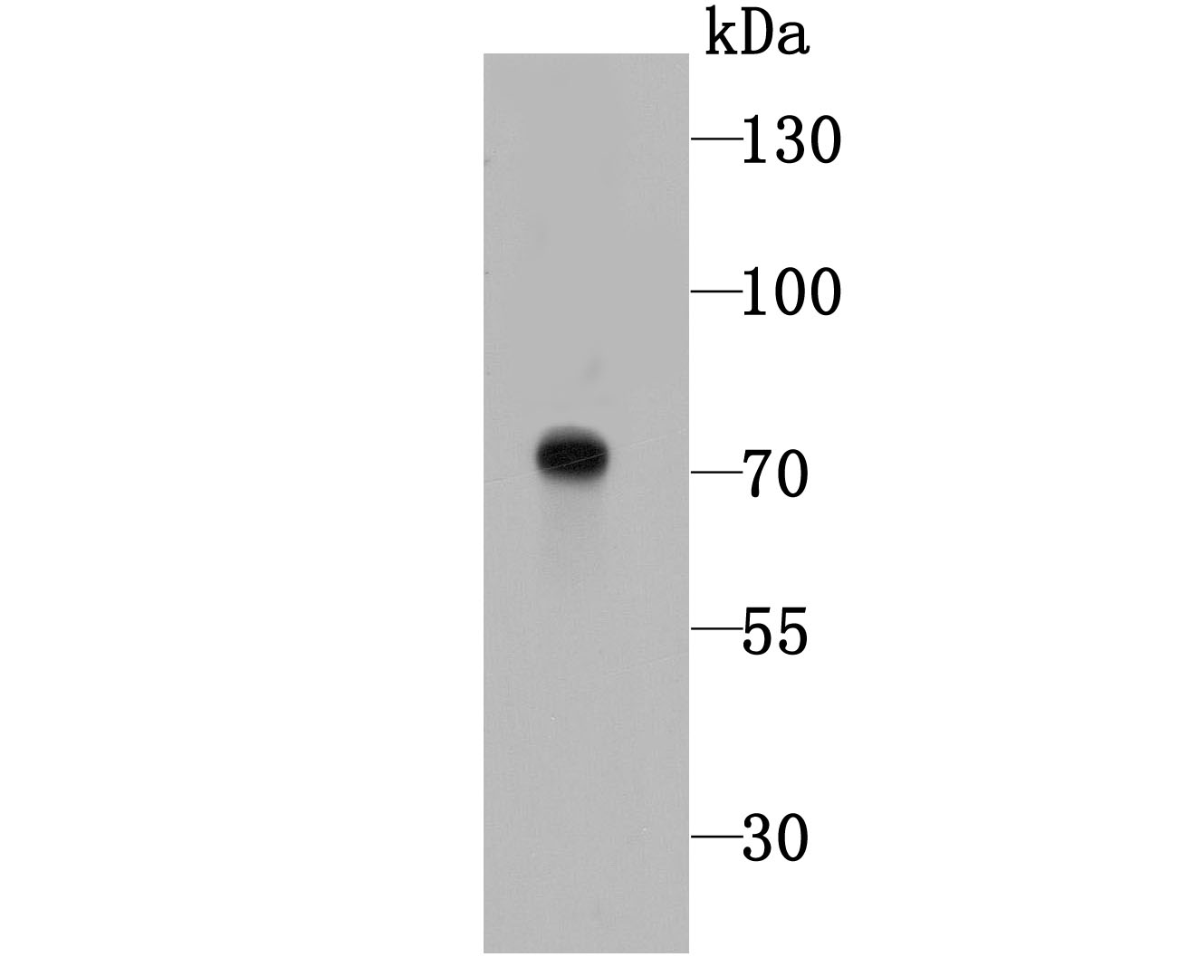
Fig1: Western blot analysis of Tyrosinase on B16F1 cell lysates using anti- Tyrosinase antibody at 1/1,000 dilution.
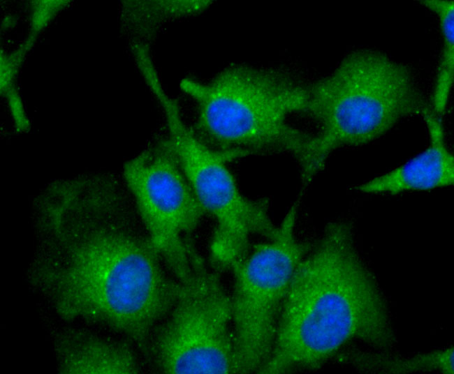
Fig2: ICC staining Tyrosinase in B16F1 cells (red). The nuclear counter stain is DAPI (blue). Cells were fixed in paraformaldehyde, permeabilised with 0.25% Triton X100/PBS.
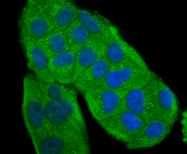
Fig3: ICC staining Tyrosinase in Hela cells (red). The nuclear counter stain is DAPI (blue). Cells were fixed in paraformaldehyde, permeabilised with 0.25% Triton X100/PBS.
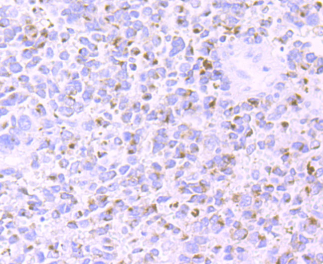
Fig4: Immunohistochemical analysis of paraffin-embedded human urethral melanoma tissue using anti-Tyrosinase antibody. Counter stained with hematoxylin.
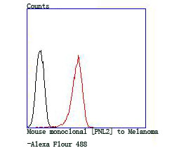
Fig5: Flow cytometric analysis of A431 cells with Tyrosinase antibody at 1/100 dilution (green) compared with an unlabelled control (cells without incubation with primary antibody; red).
Product Profile
| Product Name | Tyrosinase |
|---|---|
| Antibody Type | Primary Antibodies |
| Product description |
|
| Immunogen | Peptide |
Key Feature
| Clonality | Polyclonal |
|---|---|
| Isotype | IgG |
| Host Species | Rabbit |
| Tested Applications | |
WB:1:200-1:500 ICC:1:50-1:200 IHC:1:50-1:200 FC:1:50-1:200: |
|
| Species Reactivity | |
| Concentration | 1 mg/mL. |
Target Information
| Alternative Names | ATN antibody
CMM8 antibody
LB24 AB antibody
LB24-AB antibody
Monophenol monooxygenase antibody
OCA1 antibody
OCA1A antibody
OCAIA antibody
Oculocutaneous albinism IA antibody
SHEP3 antibody
SK29 AB antibody
SK29-AB antibody
Tumor rejection antigen AB antibody
TYR antibody
TYRO_HUMAN antibody
tyrosinase (oculocutaneous albinism IA) antibody
Tyrosinase antibody
|
|---|---|
| Molecular Weight(MW) | 70 kDa |
| Cellular Localization | Membrane. |
Database Links
| SwissProt ID | P14679 |
|---|
Application
-

Application
Fig1: Western blot analysis of Tyrosinase on B16F1 cell lysates using anti- Tyrosinase antibody at 1/1,000 dilution.
-

Application
Fig2: ICC staining Tyrosinase in B16F1 cells (red). The nuclear counter stain is DAPI (blue). Cells were fixed in paraformaldehyde, permeabilised with 0.25% Triton X100/PBS.
-

Application
Fig3: ICC staining Tyrosinase in Hela cells (red). The nuclear counter stain is DAPI (blue). Cells were fixed in paraformaldehyde, permeabilised with 0.25% Triton X100/PBS.
-

Application
Fig4: Immunohistochemical analysis of paraffin-embedded human urethral melanoma tissue using anti-Tyrosinase antibody. Counter stained with hematoxylin.
-

Application
Fig5: Flow cytometric analysis of A431 cells with Tyrosinase antibody at 1/100 dilution (green) compared with an unlabelled control (cells without incubation with primary antibody; red).
| Positive Control | B16F1, Hela, human urethral melanoma tissue, A431. |
|---|---|
| Application Notes | WB:1:200-1:500 ICC:1:50-1:200 IHC:1:50-1:200 FC:1:50-1:200: |
Additional Information
| Form | Liquid |
|---|---|
| Storage Instructions | Store at +4℃ after thawing. Aliquot store at -20℃ or -80℃. Avoid repeated freeze / thaw cycles. |
| Storage Buffer | 1*TBS (pH7.4), 0.5%BSA, 50%Glycerol. Preservative: 0.05% Sodium Azide. |
- Related products
- Camel Growth Hormone (GH) ELISA Kit OM642709
- Camel Insulin-like growth factors 1 (IGF-1) ELISA Kit OM642708
- Human HIV-1 p24 core protein (HIV-1 p24) antibody ELISA Kit OM642706
- Antigen Repair 20X pH6.0 OM642703
- 647 labeled Tyramide OM642699
-
- ASSAY KITS
-
- SERUM
2013 © Omnimabs , All Rights Reserved.

