

Immunohistochemical
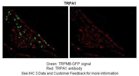
Section of TRPM8-GFP transgenic mouse trigeminal gangliaPrimary Antibody Dilution :1:250Secondary Antibody :Goat Anti-Rabbit Alexa-Fluor 594Secondary Antibody Dilution :1:1000Color/Signal Descriptions :TRPA1: Red TRPM8-GFP:: GreenGene Name :TRPA1Submitted by :Dr. Daniel McCoy, USCImmunohistochemistry
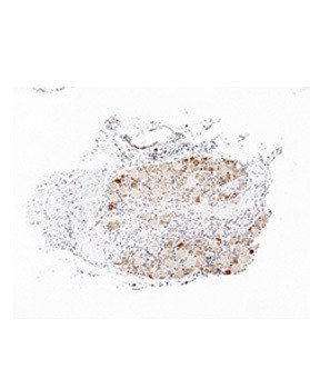
Immunohistochemistry with rat sensory ganglia tissueWestern blot
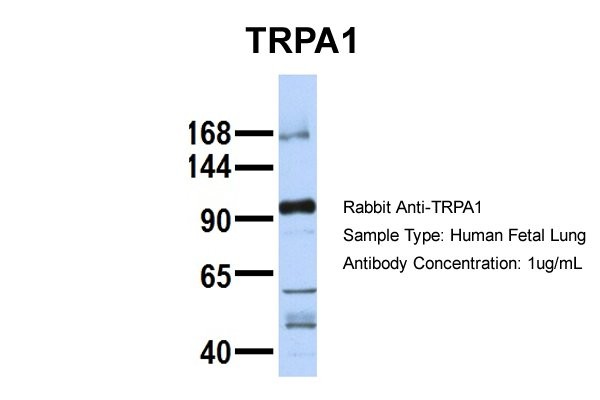
Host: Rabbit Target Name: TRPA1 Sample Tissue: Human Fetal Lung Antibody Dilution: 1.0ug/mlWestern blot
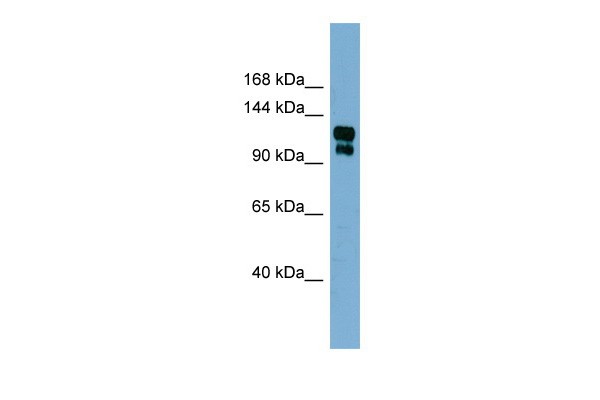
0.2-1 ug/mlELISA Titer: 1:312500Positive Control: Human StomachIF
a The co-localization of P2X7R and TRPA1 was detected using immunofluorescence in THP-1-derived macrophage. TRPA1-AF488 (green) and P2X7R-AF594 (red) were shown. Scale bar, 10 μm.c The three-dimensional image was detected with confocal Z-stack (12 planes, 1.386 μm step width) in THP-1-derived macrophage. d The co-localization of P2X7R and TRPA1 was detected using immunofluorescence in HEK293T cell line. The plasmids of rat P2X7R and rat TRPA1 were transfected transiently into HEK293T cell line. TRPA1-AF488 (green), P2X7R-AF594 (red) and DAPI (blue) were shown.WB
b Western blot analysis of TRPA1 in THP-1-derived macrophages. α-Tubulin was used as a control.IF
c Immunofluorescence staining of cytoplasmic TRPA1 in THP-1-derived macrophages. DAPI (blue) and TRPA1-AF488 (Fang et al. 2011) are shown. The scale bar represents 20 μm. d Immunofluorescence staining of TRPA1 in the membrane in THP-1-derived macrophages. The scale bar represents 50 μm.| Product Name | Rabbit anti-TRPA1 polyclonal antibody - middle region |
|---|---|
| Antibody Type | Primary Antibodies |
| Immunogen | The immunogen for anti-TRPA1 antibody: synthetic peptide directed towards the middle region of human TRPA1 |
| Clonality | Polyclonal |
|---|---|
| Isotype | IgG |
| Host Species | Rabbit |
| Tested Applications | IFIHCWB |
| WB:1:500~1:2000 IHC:1:50~1:200 IF:1:200~1:500 Notes:Optimal dilutions/concentrations should be determined by the researcher. |
|
| Species Reactivity | BovineDogGuinea PigHorseHumanMousePigRabbitRat |
| Concentration | 1mg/ml |
| Purification | Affinity purified |
| Gene Symbol | TRPA1 |
|---|---|
| Gene Synonyms | ANKTM1 |
| Gene Full Name | Transient receptor potential cation channel, subfamily A, member 1 |
| Gene Summary | The structure of the protein encoded by this gene is highly related to both the protein ankyrin and transmembrane proteins. The specific function of this protein has not yet been determined; however, studies indicate the function may involve a role in sig |
| Alternative Names | ANKTM1 |
| Molecular Weight(MW) | 127kDa |
| Sequence | 1119 amino acids |
Immunohistochemical
Section of TRPM8-GFP transgenic mouse trigeminal gangliaPrimary Antibody Dilution :1:250Secondary Antibody :Goat Anti-Rabbit Alexa-Fluor 594Secondary Antibody Dilution :1:1000Color/Signal Descriptions :TRPA1: Red TRPM8-GFP:: GreenGene Name :TRPA1Submitted by :Dr. Daniel McCoy, USC
Immunohistochemistry
Immunohistochemistry with rat sensory ganglia tissue
Western blot
Host: Rabbit Target Name: TRPA1 Sample Tissue: Human Fetal Lung Antibody Dilution: 1.0ug/ml
Western blot
0.2-1 ug/mlELISA Titer: 1:312500Positive Control: Human Stomach
IF
a The co-localization of P2X7R and TRPA1 was detected using immunofluorescence in THP-1-derived macrophage. TRPA1-AF488 (green) and P2X7R-AF594 (red) were shown. Scale bar, 10 μm.c The three-dimensional image was detected with confocal Z-stack (12 planes, 1.386 μm step width) in THP-1-derived macrophage. d The co-localization of P2X7R and TRPA1 was detected using immunofluorescence in HEK293T cell line. The plasmids of rat P2X7R and rat TRPA1 were transfected transiently into HEK293T cell line. TRPA1-AF488 (green), P2X7R-AF594 (red) and DAPI (blue) were shown.
WB
b Western blot analysis of TRPA1 in THP-1-derived macrophages. α-Tubulin was used as a control.
IF
c Immunofluorescence staining of cytoplasmic TRPA1 in THP-1-derived macrophages. DAPI (blue) and TRPA1-AF488 (Fang et al. 2011) are shown. The scale bar represents 20 μm. d Immunofluorescence staining of TRPA1 in the membrane in THP-1-derived macrophages. The scale bar represents 50 μm.| Application Notes | WB:1:500~1:2000 IHC:1:50~1:200 IF:1:200~1:500 Notes:Optimal dilutions/concentrations should be determined by the researcher. |
|---|
| Form | Liquid |
|---|---|
| Storage Instructions | Aliquot and store at -20°C. Avoid repeated freeze / thaw cycles |
| Storage Buffer | phosphate buffered saline , pH 7.4, 150mM NaCl, 0.02% sodium azide and 50% glycerol. |


