WB
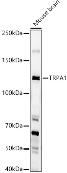
Western blot analysis of lysates from Mouse brain, using TRPA1 Rabbit pAb at 1:1000 dilution. Secondary antibody: HRP-conjugated Goat anti-Rabbit IgG (H+L)at 1:10000 dilution. Lysates/proteins: 25μg per lane. Blocking buffer: 3% nonfat dry milk in TBST. Detection: ECL Basic Kit. Exposure time: 60s.IHC
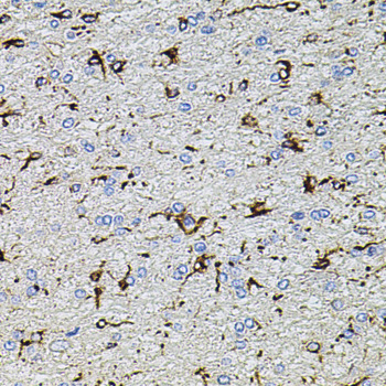
Immunohistochemistry analysis of paraffin-embedded Rat brain using TRPA1 Rabbit pAb at dilution of 1:100 (40x lens). Microwave antigen retrieval performed with 0.01M PBS Buffer (pH 7.2) prior to IHC staining.IHC
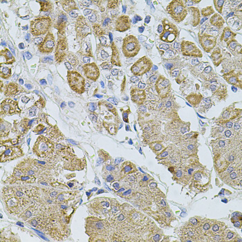
Immunohistochemistry analysis of paraffin-embedded Human stomach using TRPA1 Rabbit pAb at dilution of 1:100 (40x lens). Microwave antigen retrieval performed with 0.01M PBS Buffer (pH 7.2) prior to IHC staining.IHC
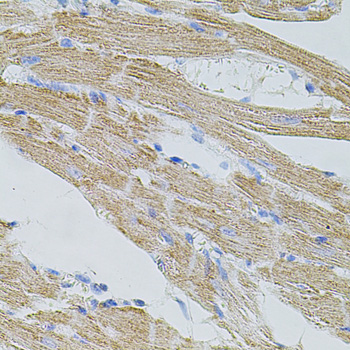
Immunohistochemistry analysis of paraffin-embedded Mouse heart using TRPA1 Rabbit pAb at dilution of 1:100 (40x lens). Microwave antigen retrieval performed with 0.01M PBS Buffer (pH 7.2) prior to IHC staining.IF-P
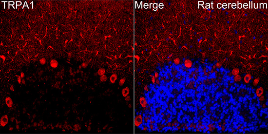
Immunofluorescence analysis of paraffin-embedded Rat cerebellum tissue using TRPA1 Rabbit pAb at a dilution of 1:200 (40x lens). Secondary antibody:Cy3 Goat Anti-Rabbit IgG (H+L) at 1:500 dilution. Blue: DAPI for nuclear staining. Perform microwave antigen retrieval with 0.01 M citrate buffer (pH 6.0) prior to IF staining.| Product Name | TRPA1 Rabbit pAb |
|---|---|
| Antibody Type | Primary Antibodies |
| Immunogen | Synthetic peptide. This information is considered to be commercially sensitive. |
| Clonality | Polyclonal |
|---|---|
| Isotype | IgG |
| Host Species | Rabbit |
| Tested Applications | IF-PIHCWB |
| WB:1:500-1:1000 IHC:1:50-1:200 IF-P:1:50-1:200 |
|
| Species Reactivity | HumanMouseRat |
| Concentration | 1mg/ml |
| Purification | Affinity purified |
| Gene Symbol | TRPA1 |
|---|---|
| Gene Synonyms | FEPS p120 FEPS1 ANKTM1 |
| Gene Full Name | transient receptor potential cation channel subfamily A member 1 |
| Gene Summary | The structure of the protein encoded by this gene is highly related to both the protein ankyrin and transmembrane proteins. The specific function of this protein has not yet been determined; however, studies indicate the function may involve a role in signal transduction and growth control. [provided by RefSeq, Jul 2008] |
| Alternative Names | FEPS p120 FEPS1 ANKTM1 TRPA1 |
| Molecular Weight(MW) | 128 kDa |
| Cellular Localization | Cell membrane, Multi-pass membrane protein. |
WB
Western blot analysis of lysates from Mouse brain, using TRPA1 Rabbit pAb at 1:1000 dilution. Secondary antibody: HRP-conjugated Goat anti-Rabbit IgG (H+L)at 1:10000 dilution. Lysates/proteins: 25μg per lane. Blocking buffer: 3% nonfat dry milk in TBST. Detection: ECL Basic Kit. Exposure time: 60s.
IHC
Immunohistochemistry analysis of paraffin-embedded Rat brain using TRPA1 Rabbit pAb at dilution of 1:100 (40x lens). Microwave antigen retrieval performed with 0.01M PBS Buffer (pH 7.2) prior to IHC staining.
IHC
Immunohistochemistry analysis of paraffin-embedded Human stomach using TRPA1 Rabbit pAb at dilution of 1:100 (40x lens). Microwave antigen retrieval performed with 0.01M PBS Buffer (pH 7.2) prior to IHC staining.
IHC
Immunohistochemistry analysis of paraffin-embedded Mouse heart using TRPA1 Rabbit pAb at dilution of 1:100 (40x lens). Microwave antigen retrieval performed with 0.01M PBS Buffer (pH 7.2) prior to IHC staining.
IF-P
Immunofluorescence analysis of paraffin-embedded Rat cerebellum tissue using TRPA1 Rabbit pAb at a dilution of 1:200 (40x lens). Secondary antibody:Cy3 Goat Anti-Rabbit IgG (H+L) at 1:500 dilution. Blue: DAPI for nuclear staining. Perform microwave antigen retrieval with 0.01 M citrate buffer (pH 6.0) prior to IF staining.| Application Notes | WB:1:500-1:1000 IHC:1:50-1:200 IF-P:1:50-1:200 |
|---|
| Form | Liquid |
|---|---|
| Storage Instructions | Store at -20℃. Avoid freeze / thaw cycles. |
| Storage Buffer | Buffer: PBS with 0.05% proclin300, 0.05% BSA, 50% glycerol, pH7.3. |


