
Western blot
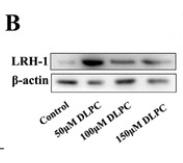
2.5ug/mlWB
(B) Western blotting of LRH-1 protein in granulosa cells by treatment with various concentrations of DLPC.IHC
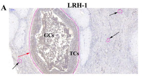
(A) Immunochemistry assay was performed to visualize the localization of LRH-1 in bovine ovarian cells. Immuno-specific staining was brown, indicating LRH-1-positive cells. Immunohistochemistry was performed on three different ovarian slides from each of three bovines. Atretic follicles were indicated with the red arrow, whereas healthy follicles were indicated with black arrows. Bar: 200 μm.| Product Name | NR5A2 antibody - N-terminal region |
|---|---|
| Antibody Type | Primary Antibodies |
| Product description |
|
| Immunogen | The immunogen for anti-NR5A2 antibody: synthetic peptide directed towards the N terminal of human NR5A2 |
| Clonality | Polyclonal |
|---|---|
| Isotype | IgG |
| Host Species | Rabbit |
| Tested Applications | IHCWB |
| Concentration | 1mg/ml |
| Purification | Protein A |
| Gene Symbol | NR5A2 |
|---|---|
| Gene Synonyms | B1F B1F2 CPF FTF FTZ-F1 FTZ-F1beta LRH-1 hB1F hB1F-2 LRH1 |
| Gene Full Name | Nuclear receptor subfamily 5, group A, member 2 |
| Gene Summary | NR5A2 binds to the sequence element 5'-AACGACCGACCTTGAG-3' of the enhancer II of hepatitis B virus genes, a critical cis-element of their expression and regulation. It may be responsable for the liver-specific activity of enhancer II, probably in combination with other hepatocyte transcription factors. It is a key regulator of cholesterol 7-alpha-hydroxylase gene (CYP7A) expression in liver. It may also contribute to the regulation of pancreas-specific genes and play important roles in embryonic development. |
| Alternative Names | B1F B1F2 CPF FTF FTZ-F1 FTZ-F1beta LRH-1 hB1F hB1F-2 LRH1 |
| Molecular Weight(MW) | 61kDa |
| Sequence | 541 amino acids |
Western blot
2.5ug/ml
WB
(B) Western blotting of LRH-1 protein in granulosa cells by treatment with various concentrations of DLPC.
IHC
(A) Immunochemistry assay was performed to visualize the localization of LRH-1 in bovine ovarian cells. Immuno-specific staining was brown, indicating LRH-1-positive cells. Immunohistochemistry was performed on three different ovarian slides from each of three bovines. Atretic follicles were indicated with the red arrow, whereas healthy follicles were indicated with black arrows. Bar: 200 μm.| Form | Liquid |
|---|---|
| Storage Instructions | Aliquot and store at -20°C. Avoid repeated freeze / thaw cycles |
| Storage Buffer | phosphate buffered saline , pH 7.4, 150mM NaCl, 0.02% sodium azide and 50% glycerol. |


