Western blot
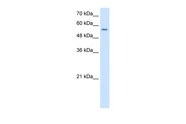
1.25ug/mlPositive Control: HepG2 cell lysateWB
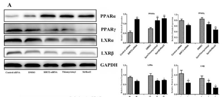
(A) SIRT2 knockdown blocked PPARγ and LXRα, promoted PPARα. The GCs were transfected with SIRT2-siRNA 4 plasmids for 48 h and treated with the SIRT2 inhibitors Thiomyristoyl (2 μM) and SirReal2 (5 μM) for 24 h. Western blotting for PPARα, PPARγ, and LXRα are shown. According to the western blot results, the protein ratios were analyzed in treatment groups relative to the GAPDH protein abundance.WB
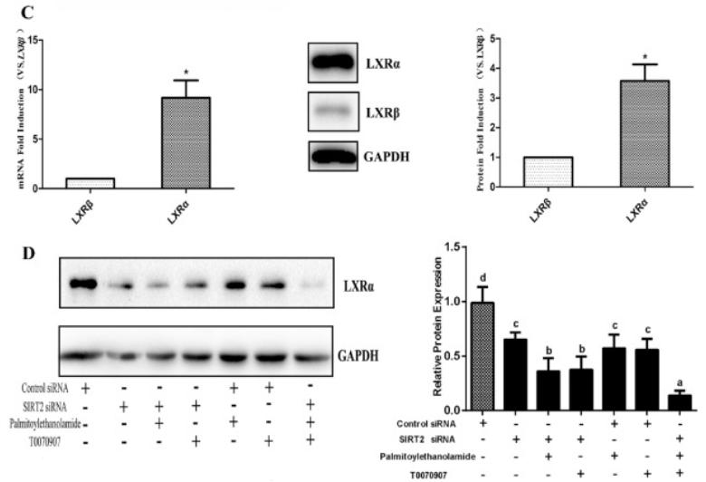
(C) LXRα predominantly expressed in GCs, but not LXRβ. The mRNA and protein abundance of LXRα and LXRβ were respectively detected by RT-qPCR and western blot in primary GCs. The expression level of LXRβ was arbitrarily set at 1.0, and the mRNA and protein expression ratios of LXRα/ LXRβ are shown. (D) SIRT2 knockdown blocked the expression of LXRα via PPARs. The primary GCs were pretreated with SIRT2-siRNA or control siRNA for 48 h, and then treated with or without the PPARα agonist Palmitoylethanolamide (20 μM) or the PPARγ antagonist T0070907 (2 μM) for 24 h. The LXRα protein abundance was measured by western blotting. The protein ratios were analyzed in treatment groups relative to the GAPDH protein abundance.| Product Name | Rabbit anti-NR1H2 polyclonal antibody - middle region |
|---|---|
| Antibody Type | Primary Antibodies |
| Product description |
|
| Immunogen | The immunogen for anti-NR1H2 antibody: synthetic peptide directed towards the middle region of human NR1H2 |
| Clonality | Polyclonal |
|---|---|
| Isotype | IgG |
| Host Species | Rabbit |
| Tested Applications | WB |
| WB:1:500~1:2000 Notes:Optimal dilutions/concentrations should be determined by the researcher. |
|
| Species Reactivity | BovineDogGuinea PigHorseHumanMousePigRabbitRat |
| Concentration | 1mg/ml |
| Purification | Protein A |
| Gene Symbol | NR1H2 |
|---|---|
| Gene Synonyms | LXR-b LXRB NER NER-I RIP15 UNR |
| Gene Full Name | Nuclear receptor subfamily 1, group H, member 2 |
| Gene Summary | The LX receptors (LXRs) were originally identified as orphan members of the nuclear receptor superfamily because their ligands were unknown. Like other receptors in the family, LXRs heterodimerize with retinoid X receptor and bind to specific response elements (LXREs) characterized by direct repeats separated by 4 nucleotides. Two genes, alpha (LXRA) and beta, are known to encode LXR proteins.The LX receptors (LXRs) were originally identified as orphan members of the nuclear receptor superfamily because their ligands were unknown. Like other receptors in the family, LXRs heterodimerize with retinoid X receptor (see MIM 180245) and bind to specific response elements (LXREs) characterized by direct repeats separated by 4 nucleotides. Two genes, alpha (LXRA, MIM 602423) and beta, are known to encode LXR proteins.The LX receptors (LXRs) were originally identified as orphan members of the nuclear receptor superfamily because their ligands were unknown. Like other receptors in the family, LXRs heterodimerize with retinoid X receptor (see MIM 180245) and bind to specific response elements (LXREs) characterized by direct repeats separated by 4 nucleotides. Two genes, alpha (LXRA, MIM 602423) and beta, are known to encode LXR proteins (Song et al., 1995).[supplied by OMIM]. |
| Alternative Names | LXR-b LXRB NER NER-I RIP15 UNR |
| Molecular Weight(MW) | 51kDa |
| Sequence | 461 amino acids |
Western blot
1.25ug/mlPositive Control: HepG2 cell lysate
WB
(A) SIRT2 knockdown blocked PPARγ and LXRα, promoted PPARα. The GCs were transfected with SIRT2-siRNA 4 plasmids for 48 h and treated with the SIRT2 inhibitors Thiomyristoyl (2 μM) and SirReal2 (5 μM) for 24 h. Western blotting for PPARα, PPARγ, and LXRα are shown. According to the western blot results, the protein ratios were analyzed in treatment groups relative to the GAPDH protein abundance.
WB
(C) LXRα predominantly expressed in GCs, but not LXRβ. The mRNA and protein abundance of LXRα and LXRβ were respectively detected by RT-qPCR and western blot in primary GCs. The expression level of LXRβ was arbitrarily set at 1.0, and the mRNA and protein expression ratios of LXRα/ LXRβ are shown. (D) SIRT2 knockdown blocked the expression of LXRα via PPARs. The primary GCs were pretreated with SIRT2-siRNA or control siRNA for 48 h, and then treated with or without the PPARα agonist Palmitoylethanolamide (20 μM) or the PPARγ antagonist T0070907 (2 μM) for 24 h. The LXRα protein abundance was measured by western blotting. The protein ratios were analyzed in treatment groups relative to the GAPDH protein abundance.| Application Notes | WB:1:500~1:2000 Notes:Optimal dilutions/concentrations should be determined by the researcher. |
|---|
| Form | Liquid |
|---|---|
| Storage Instructions | Aliquot and store at -20°C. Avoid repeated freeze / thaw cycles |
| Storage Buffer | phosphate buffered saline , pH 7.4, 150mM NaCl, 0.02% sodium azide and 50% glycerol. |


