WB
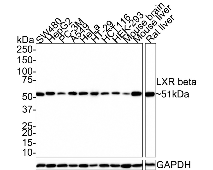
Western blot analysis of LXR beta on different lysates with Rabbit anti-LXR beta antibody at 1/1,000 dilution. Lane 1: SW480 cell lysate (20 µg/Lane), Lane 2: HepG2 cell lysate (20 µg/Lane), Lane 3: PC-3M cell lysate (20 µg/Lane), Lane 4: A549 cell lysate (20 µg/Lane), Lane 5: HeLa cell lysate (20 µg/Lane), Lane 6: HT-29 cell lysate (20 µg/Lane), Lane 7: HCT116 cell lysate (20 µg/Lane), Lane 8: HEK-293 cell lysate (20 µg/Lane), Lane 9: Mouse brain tissue lysate (40 µg/Lane), Lane 10: Mouse liver tissue lysate (40 µg/Lane), Lane 11: Rat liver tissue lysate (40 µg/Lane), Exposure time: 2 minutes; 4-20% SDS-PAGE gel. Proteins were transferred to a PVDF membrane and blocked with 5% NFDM/TBST for 1 hour at room temperature. The primary antibody at 1/1,000 dilution was used in 5% NFDM/TBST at room temperature for 2 hours. Goat Anti-Rabbit IgG - HRP Secondary Antibody at 1:100,000 dilution was used for 1 hour at room temperature.IICC/IF
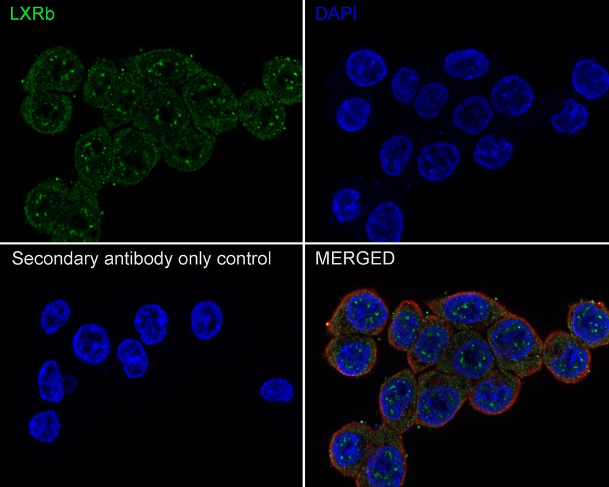
Immunocytochemistry analysis of HCT116 cells labeling LXR beta with Rabbit anti-LXR beta antibody at 1/100 dilution. Cells were fixed in 4% paraformaldehyde for 10 minutes at 37 ℃, permeabilized with 0.05% Triton X-100 in PBS for 20 minutes, and then blocked with 2% negative goat serum for 30 minutes at room temperature. Cells were then incubated with Rabbit anti-LXR beta antibody at 1/100 dilution in 2% negative goat serum overnight at 4 ℃. Goat Anti-Rabbit IgG H&L (488) was used as the secondary antibody at 1/1,000 dilution. Nuclear DNA was labelled in blue with DAPI. Beta tubulin (red) was stained at 1/200 dilution overnight at +4℃. Goat Anti-Mouse IgG H&L (594) was used as the secondary antibody at 1/1,000 dilution.Immunocytochemistry analysis of HCT116 cells labeling LXR beta with Rabbit anti-LXR beta antibody at 1/100 dilution. Cells were fixed in 4% paraformaldehyde for 10 minutes at 37 ℃, permeabilized with 0.05% Triton X-100 in PBS for 20 minutes, and then blocked with 2% negative goat serum for 30 minutes at room temperature. Cells were then incubated with Rabbit anti-LXR beta antibody at 1/100 dilution in 2% negative goat serum overnight at 4 ℃. Goat Anti-Rabbit IgG H&L (488) was used as the secondary antibody at 1/1,000 dilution. Nuclear DNA was labelled in blue with DAPI. Beta tubulin (red) was stained at 1/200 dilution overnight at +4℃. Goat Anti-Mouse IgG H&L (594) was used as the secondary antibody at 1/1,000 dilution.| Product Name | LXR beta Recombinant Rabbit Monoclonal Antibody |
|---|---|
| Antibody Type | Primary Antibodies |
| Immunogen | Synthetic peptide within human LXR beta aa 50-100/460. |
| Clonality | monoclonal |
|---|---|
| Isotype | IgG |
| Host Species | Rabbit |
| Tested Applications | ICC/IFWB |
| WB:1:1000 ICC/IF:1:100 |
|
| Species Reactivity | HumanMouseRat |
| Concentration | 1mg/ml |
| Purification | Protein A |
| Gene Symbol | NR1H2 |
|---|---|
| Gene Synonyms | NER UNR LXRB LXR-b NER-I RIP15 |
| Gene Full Name | nuclear receptor subfamily 1 group H member 2 |
| Gene Summary | The liver X receptors, LXRA (NR1H3; MIM 602423) and LXRB, form a subfamily of the nuclear receptor superfamily and are key regulators of macrophage function, controlling transcriptional programs involved in lipid homeostasis and inflammation. The inducible LXRA is highly expressed in liver, adrenal gland, intestine, adipose tissue, macrophages, lung, and kidney, whereas LXRB is ubiquitously expressed. Ligand-activated LXRs form obligate heterodimers with retinoid X receptors (RXRs; see MIM 180245) and regulate expression of target genes containing LXR response elements (summary by Korf et al., 2009 [PubMed 19436111]).[supplied by OMIM, Jan 2010] |
| Alternative Names | Liver X nuclear receptor beta antibody Liver X receptor beta antibody LX receptor beta antibody LXR b antibody LXR beta antibody LXRB antibody NER antibody NER I antibody NER1 antibody NR1H 2 antibody Liver X nuclear receptor beta antibody Liver X receptor beta antibody LX receptor beta antibody LXR b antibody LXR beta antibody LXRB antibody NER antibody NER I antibody NER1 antibody NR1H 2 antibody NR1H2 antibody NR1H2_HUMAN antibody Nuclear orphan receptor LXR beta antibody Nuclear receptor NER antibody Nuclear receptor subfamily 1 group H member 2 antibody OR-1 antibody Oxysterols receptor LXR-beta antibody RIP15 antibody Steroid hormone nuclear receptor NER antibody Ubiquitously-expressed nuclear receptor antibody UNR antibody |
| Molecular Weight(MW) | 51kDa |
| Cellular Localization | Nucleus. |
WB
Western blot analysis of LXR beta on different lysates with Rabbit anti-LXR beta antibody at 1/1,000 dilution. Lane 1: SW480 cell lysate (20 µg/Lane), Lane 2: HepG2 cell lysate (20 µg/Lane), Lane 3: PC-3M cell lysate (20 µg/Lane), Lane 4: A549 cell lysate (20 µg/Lane), Lane 5: HeLa cell lysate (20 µg/Lane), Lane 6: HT-29 cell lysate (20 µg/Lane), Lane 7: HCT116 cell lysate (20 µg/Lane), Lane 8: HEK-293 cell lysate (20 µg/Lane), Lane 9: Mouse brain tissue lysate (40 µg/Lane), Lane 10: Mouse liver tissue lysate (40 µg/Lane), Lane 11: Rat liver tissue lysate (40 µg/Lane), Exposure time: 2 minutes; 4-20% SDS-PAGE gel. Proteins were transferred to a PVDF membrane and blocked with 5% NFDM/TBST for 1 hour at room temperature. The primary antibody at 1/1,000 dilution was used in 5% NFDM/TBST at room temperature for 2 hours. Goat Anti-Rabbit IgG - HRP Secondary Antibody at 1:100,000 dilution was used for 1 hour at room temperature.
IICC/IF
Immunocytochemistry analysis of HCT116 cells labeling LXR beta with Rabbit anti-LXR beta antibody at 1/100 dilution. Cells were fixed in 4% paraformaldehyde for 10 minutes at 37 ℃, permeabilized with 0.05% Triton X-100 in PBS for 20 minutes, and then blocked with 2% negative goat serum for 30 minutes at room temperature. Cells were then incubated with Rabbit anti-LXR beta antibody at 1/100 dilution in 2% negative goat serum overnight at 4 ℃. Goat Anti-Rabbit IgG H&L (488) was used as the secondary antibody at 1/1,000 dilution. Nuclear DNA was labelled in blue with DAPI. Beta tubulin (red) was stained at 1/200 dilution overnight at +4℃. Goat Anti-Mouse IgG H&L (594) was used as the secondary antibody at 1/1,000 dilution.Immunocytochemistry analysis of HCT116 cells labeling LXR beta with Rabbit anti-LXR beta antibody at 1/100 dilution. Cells were fixed in 4% paraformaldehyde for 10 minutes at 37 ℃, permeabilized with 0.05% Triton X-100 in PBS for 20 minutes, and then blocked with 2% negative goat serum for 30 minutes at room temperature. Cells were then incubated with Rabbit anti-LXR beta antibody at 1/100 dilution in 2% negative goat serum overnight at 4 ℃. Goat Anti-Rabbit IgG H&L (488) was used as the secondary antibody at 1/1,000 dilution. Nuclear DNA was labelled in blue with DAPI. Beta tubulin (red) was stained at 1/200 dilution overnight at +4℃. Goat Anti-Mouse IgG H&L (594) was used as the secondary antibody at 1/1,000 dilution.| Application Notes | WB:1:1000 ICC/IF:1:100 |
|---|
| Form | Liquid |
|---|---|
| Storage Instructions | Store at +4℃ after thawing. Aliquot store at -20℃. Avoid repeated freeze / thaw cycles. |
| Storage Buffer | 1*TBS (pH7.4), 0.05% BSA, 40% Glycerol. Preservative: 0.05% Sodium Azide. |


