Immunohistochemical
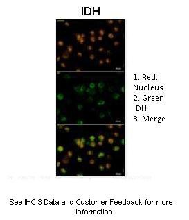
Human glioma cellsPrimary Antibody Dilution :1:200Secondary Antibody :Anti-rabbit-GFPSecondary Antibody Dilution :1:5000Color/Signal Descriptions :1. Red: Nucleus 2. Green: IDH 3. MergeGene Name :IDH1Submitted by :ELIAS EL HABR, PhD, Post-Doctoral fellow, INSERM U894 Glial Plasticity Team, Psychiatry& Neurosciences Center, St Anne Hospital, 2 Ter, Rue d'Alesia, 75014 ParisWestern blot
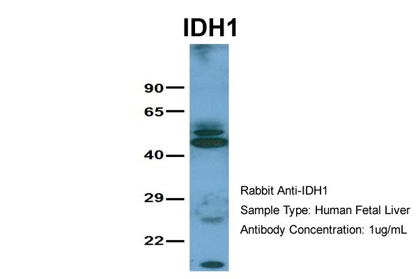
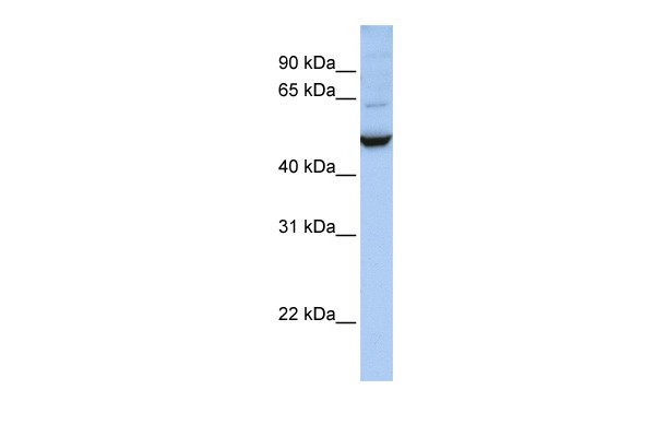
Host: Rabbit Target Name: IDH1 Sample Tissue: Human Fetal Liver Antibody Dilution: 1.0ug/mlWestern blot
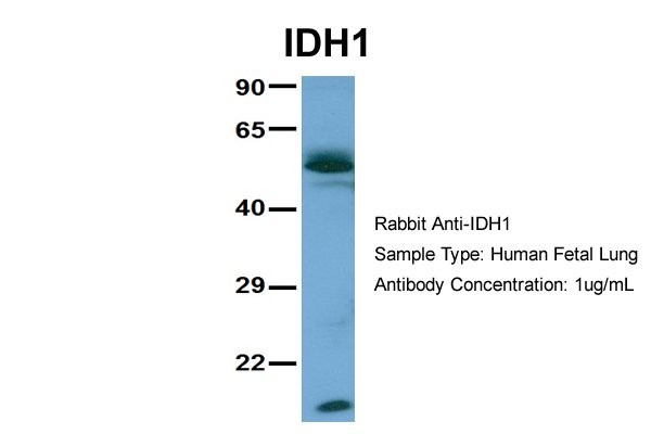
Host: Rabbit Target Name: IDH1 Sample Tissue: Human Fetal Lung Antibody Dilution: 1.0ug/mlWestern blot
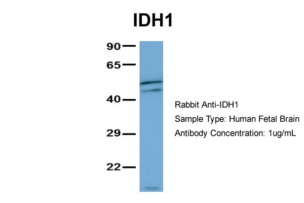
Host: Rabbit Target Name: IDH1 Sample Tissue: Human Fetal Brain Antibody Dilution: 1.0ug/mlWestern blot
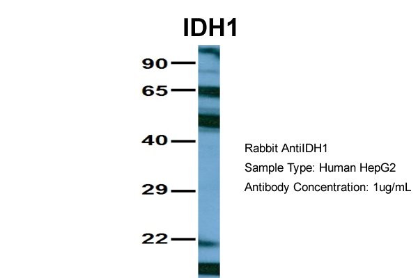
Host: Rabbit Target Name: IDH1 Sample Tissue: HepG2 Antibody Dilution: 1.0ug/mlWestern blot
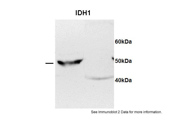
IDH1 antibody - C-terminal region validated by WB using HEPG2 lysate + Drosophila extract at 1:1000.Western blot
Sample type: 1. HEPG2 (50ug)2. Drosophila extract (50ug)Primary dilution: 1:1000Secondary Antibody: mouse anti-Rabbit HRPSecondary Dilution: 1:5000Image Submitted by: Xinyang BingLaurentian University| Product Name | Rabbit anti-IDH1 polyclonal antibody - C-terminal region |
|---|---|
| Antibody Type | Primary Antibodies |
| Antigen Alias | IDH, IDP, PICD, IDCD, IDPC |
| Immunogen | The immunogen for anti-IDH1 antibody: synthetic peptide directed towards the C terminal of human IDH1 |
| Clonality | Polyclonal |
|---|---|
| Isotype | IgG |
| Host Species | Rabbit |
| Tested Applications | WBIHC |
| WB:1:500~1:2000 IHC:1:50~1:200 Notes:Optimal dilutions/concentrations should be determined by the researcher. |
|
| Species Reactivity | BovineDogGuinea PigHorseHumanMousePigRabbitRat |
| Concentration | 1mg/ml |
| Purification | Affinity purified |
| Gene Symbol | IDH1 |
|---|---|
| Gene Synonyms | IDH IDP PICD IDCD IDPC |
| Gene Full Name | Isocitrate dehydrogenase 1 (NADP+), soluble |
| Gene Summary | Isocitrate dehydrogenases catalyze the oxidative decarboxylation of isocitrate to 2-oxoglutarate. These enzymes belong to two distinct subclasses, one of which utilizes NAD(+) as the electron acceptor and the other NADP(+).The protein encoded by this gene is the NADP(+)-dependent isocitrate dehydrogenase found in the cytoplasm and peroxisomes. It contains the PTS-1 peroxisomal targeting signal sequence. The presence of this enzyme in peroxisomes suggests roles in the regeneration of NADPH for intraperoxisomal reductions, such as the conversion of 2, 4-dienoyl-CoAs to 3-enoyl-CoAs, as well as in peroxisomal reactions that consume 2-oxoglutarate, namely the alpha-hydroxylation of phytanic acid. The cytoplasmic enzyme serves a significant role in cytoplasmic NADPH production.Isocitrate dehydrogenases catalyze the oxidative decarboxylation of isocitrate to 2-oxoglutarate. These enzymes belong to two distinct subclasses, one of which utilizes NAD(+) as the electron acceptor and the other NADP(+). Five isocitrate dehydrogenases have been reported: three NAD(+)-dependent isocitrate dehydrogenases, which localize to the mitochondrial matrix, and two NADP(+)-dependent isocitrate dehydrogenases, one of which is mitochondrial and the other predominantly cytosolic. Each NADP(+)-dependent isozyme is a homodimer. The protein encoded by this gene is the NADP(+)-dependent isocitrate dehydrogenase found in the cytoplasm and peroxisomes. It contains the PTS-1 peroxisomal targeting signal sequence. The presence of this enzyme in peroxisomes suggests roles in the regeneration of NADPH for intraperoxisomal reductions, such as the conversion of 2, 4-dienoyl-CoAs to 3-enoyl-CoAs, as well as in peroxisomal reactions that consume 2-oxoglutarate, namely the alpha-hydroxylation of phytanic acid. The cytoplasmic enzyme serves a significant role in cytoplasmic NADPH production. Publication Note: This RefSeq record includes a subset of the publications that are available for this gene. Please see the Entrez Gene record to access additional publications. |
| Alternative Names | IDH IDP PICD IDCD IDPC |
| Molecular Weight(MW) | 47kDa |
| Sequence | 414 amino acids |
Immunohistochemical
Human glioma cellsPrimary Antibody Dilution :1:200Secondary Antibody :Anti-rabbit-GFPSecondary Antibody Dilution :1:5000Color/Signal Descriptions :1. Red: Nucleus 2. Green: IDH 3. MergeGene Name :IDH1Submitted by :ELIAS EL HABR, PhD, Post-Doctoral fellow, INSERM U894 Glial Plasticity Team, Psychiatry& Neurosciences Center, St Anne Hospital, 2 Ter, Rue d'Alesia, 75014 Paris
Western blot
Host: Rabbit Target Name: IDH1 Sample Tissue: Human Fetal Liver Antibody Dilution: 1.0ug/ml
Western blot
Host: Rabbit Target Name: IDH1 Sample Tissue: Human Fetal Lung Antibody Dilution: 1.0ug/ml
Western blot
Host: Rabbit Target Name: IDH1 Sample Tissue: Human Fetal Brain Antibody Dilution: 1.0ug/ml
Western blot
Host: Rabbit Target Name: IDH1 Sample Tissue: HepG2 Antibody Dilution: 1.0ug/ml
Western blot
IDH1 antibody - C-terminal region validated by WB using HEPG2 lysate + Drosophila extract at 1:1000.
Western blot
Sample type: 1. HEPG2 (50ug)2. Drosophila extract (50ug)Primary dilution: 1:1000Secondary Antibody: mouse anti-Rabbit HRPSecondary Dilution: 1:5000Image Submitted by: Xinyang BingLaurentian University| Application Notes | WB:1:500~1:2000 IHC:1:50~1:200 Notes:Optimal dilutions/concentrations should be determined by the researcher. |
|---|
| Form | Liquid |
|---|---|
| Storage Instructions | Aliquot and store at -20°C. Avoid repeated freeze / thaw cycles |
| Storage Buffer | phosphate buffered saline , pH 7.4, 150mM NaCl, 0.02% sodium azide and 50% glycerol. |


