Application
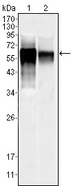
Figure 1: Western blot analysis using anti-MAPK10 mAb against NIH/3T3 (1) and SKN-SH (2) cell lysate.Application
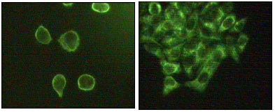
Figure 2: Immunofluorescence staining of methanol-fixed A431(left) and Hela(right) cells showing cytoplasmic and membrane localization.| Product Name | MAPK10 Antibody |
|---|---|
| Antibody Type | Primary Antibodies |
| Antigen Alias | JNK3; JNK3A; PRKM10; p54bSAPK |
| Clonality | Monoclonal |
|---|---|
| Isotype | IgG1 |
| Clone Number | 10E4A4 |
| Host Species | Mouse |
| Tested Applications | WBIF |
| WB:1:500 - 2000. IF |
|
| Species Reactivity | Human |
| Concentration | 1mg/ml |
| Gene Synonyms | JNK3 JNK3A PRKM10 SAPK1B |
|---|---|
| Alternative Names | JNK3 JNK3A PRKM10 p54bSAPK |
| Molecular Weight(MW) | 52585 Da |
| Function | Serine/threonine-protein kinase involved in various processes such as neuronal proliferation, differentiation, migration and programmed cell death. Extracellular stimuli such as proinflammatory cytokines or physical stress stimulate the stress- activated protein kinase/c-Jun N-terminal kinase (SAP/JNK) signaling pathway. In this cascade, two dual specificity kinases MAP2K4/MKK4 and MAP2K7/MKK7 phosphorylate and activate MAPK10/JNK3. In turn, MAPK10/JNK3 phosphorylates a number of transcription factors, primarily components of AP-1 such as JUN and ATF2 and thus regulates AP-1 transcriptional activity. Plays regulatory roles in the signaling pathways during neuronal apoptosis. Phosphorylates the neuronal microtubule regulator STMN2. Acts in the regulation of the beta-amyloid precursor protein/APP signaling during neuronal differentiation by phosphorylating APP. Participates also in neurite growth in spiral ganglion neurons |
| Tissue Specificity | Specific to a subset of neurons in the nervous system. Present in the hippocampus and areas, cerebellum, striatum, brain stem, and weakly in the spinal cord. Very weak expression in testis and kidney |
| Cellular Localization | Cytoplasm. Membrane; Lipid-anchor. Nucleus. Mitochondrion. Note=Palmitoylation regulates MAPK10 trafficking to cytoskeleton. Recruited to the mitochondria in the presence of SARM1 (By similarity) |
| Entrez Gene | 5602 |
|---|---|
| Nucleotide Accession | NP_620448.1 |
Application
Figure 1: Western blot analysis using anti-MAPK10 mAb against NIH/3T3 (1) and SKN-SH (2) cell lysate.
Application
Figure 2: Immunofluorescence staining of methanol-fixed A431(left) and Hela(right) cells showing cytoplasmic and membrane localization.| Application Notes | WB:1:500 - 2000. IF |
|---|
| Form | Liquid |
|---|---|
| Storage Instructions | For short-term storage, store at 4° C. For long-term storage, aliquot and store at -20ºC or below. Avoid multiple freeze-thaw cycles. |
| Storage Buffer | Purified recombinant fragment of human MAPK10 (aa28-233) expressed in E. Coli. |


