Application
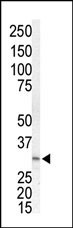
Western blot analysis of anti-MKK3 Pab in Jurkat cell lysate. MKK3 (arrow) was detected using purified Pab. Secondary HRP-anti-rabbit was used for signal visualization with chemiluminescence.Application
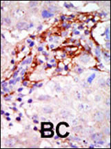
Formalin-fixed and paraffin-embedded human cancer tissue reacted with the primary antibody, which was peroxidase-conjugated to the secondary antibody, followed by AEC staining. This data demonstrates the use of this antibody for immunohistochemistry; clinical relevance has not been evaluated. BC = breast carcinoma; HC = hepatocarcinoma.Application
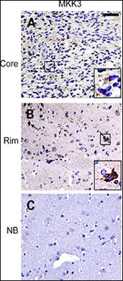
IHC of matched glioma core (top row) and rim (middle row) samples as well as normal brain control (bottom row) from a glioma invasion specific tissue microarray. Insets, individual tumor cells (black squares) are presented at higher magnification to facilitate interpretation of the staining pattern. Staining for MKK3 and pMKK3 exhibits strong signal in majority of invasive cells (B ) compared with stationary cells from tumor core (A ). Normal brain control shows only weak staining in reactive astrocytes (C ).| Product Name | MEK3 (MAP2K3) Antibody (N-term) |
|---|---|
| Antibody Type | Primary Antibodies |
| Antigen Alias | MAP2K3; MEK3; MKK3; PRKMK3; SKK2; Dual specificity mitogen-activated protein kinase kinase 3; MAPK/ERK kinase 3; Stress-activated protein kinase kinase 2 |
| Clonality | Polyclonal |
|---|---|
| Isotype | Ig |
| Host Species | Rabbit |
| Tested Applications | WBIHC |
| WB:1:100~500 IHC |
|
| Species Reactivity | Human |
| Concentration | 1mg/ml |
| Gene Synonyms | MEK3 MKK3 PRKMK3 SKK2 |
|---|---|
| Alternative Names | MAP2K3 MEK3 MKK3 PRKMK3 SKK2 Dual specificity mitogen-activated protein kinase kinase 3 MAPK/ERK kinase 3 Stress-activated protein kinase kinase 2 |
| Molecular Weight(MW) | 39318 Da |
| Function | Dual specificity kinase. Is activated by cytokines and environmental stress in vivo. Catalyzes the concomitant phosphorylation of a threonine and a tyrosine residue in the MAP kinase p38 |
| Tissue Specificity | Abundant expression is seen in the skeletal muscle. It is also widely expressed in other tissues |
| Entrez Gene | 5606 |
|---|
Application
Western blot analysis of anti-MKK3 Pab in Jurkat cell lysate. MKK3 (arrow) was detected using purified Pab. Secondary HRP-anti-rabbit was used for signal visualization with chemiluminescence.
Application
Formalin-fixed and paraffin-embedded human cancer tissue reacted with the primary antibody, which was peroxidase-conjugated to the secondary antibody, followed by AEC staining. This data demonstrates the use of this antibody for immunohistochemistry; clinical relevance has not been evaluated. BC = breast carcinoma; HC = hepatocarcinoma.
Application
IHC of matched glioma core (top row) and rim (middle row) samples as well as normal brain control (bottom row) from a glioma invasion specific tissue microarray. Insets, individual tumor cells (black squares) are presented at higher magnification to facilitate interpretation of the staining pattern. Staining for MKK3 and pMKK3 exhibits strong signal in majority of invasive cells (B ) compared with stationary cells from tumor core (A ). Normal brain control shows only weak staining in reactive astrocytes (C ).| Application Notes | WB:1:100~500 IHC |
|---|
| Form | Liquid |
|---|---|
| Storage Instructions | For short-term storage, store at 4° C. For long-term storage, aliquot and store at -20ºC or below. Avoid multiple freeze-thaw cycles. |
| Storage Buffer | Purified polyclonal antibody supplied in PBS with 0.09% (W/V) sodium azide. This antibody is purified through a protein G column, eluted with high and low pH buffers and neutralized immediately, followed by dialysis against PBS. |


